ALERT!
This site is not optimized for Internet Explorer 8 (or older).
Please upgrade to a newer version of Internet Explorer or use an alternate browser such as Chrome or Firefox.
Surgical Resection and Reconstruction of a Large Anterior Chest Dermatofibrosarcoma Protuberans
Jan A, Shah SA, Tariq M, Shah SM, Muhammad A. Surgical Resection and Reconstruction of a Large Anterior Chest Dermatofibrosarcoma Protuberans. January 2019. doi:10.25373/ctsnet.7611527.
Introduction
Dermatofibrosarcoma protuberans (DFSP) is a relatively uncommon cutaneous malignancy. Although metastasis rarely occurs, DFSP is a locally aggressive tumor with a high recurrence rate. It arises from the dermis and invades deeper tissues (eg, fat, fascia, muscle, bone) [1]. DFSP is the most common type of cutaneous sarcoma, with an incidence of 0.8 to 5 cases per million population per year. DFSP most commonly occurs on the trunk (42-72%), followed by the proximal extremities (16-30%). It rarely occurs above the neck (10-16%) [1]. Fibrosarcomatous DFSP is a more aggressive and progressive form with a higher rate of metastasis, hence labelled as fibrosarcomatous (high grade 2(G2)) [2, 3].
Case Report

Figure 1. A large anterior chest wall tumor.
A 28-year-old man who worked as a laborer presented to the authors’ outpatient clinic with a large fungating mass on the anterior chest wall. The patient gave a history of growth during the prior 6 to 8 months. Because of financial concerns, he did not seek medical care earlier. He had no significant past medical history. On examination, he had a 17 x 16 x 7.5 cm mass (Figure 1).

Figure 2. A CT scan axial section showing the chest wall tumor in the skin and subcutaneous tissue.
The authors performed preoperative staging examination and imaging. There was no evidence of tumor apart from what was on the chest wall. He had an incisional biopsy that demonstrated angiomyxoma. On the computed tomography (CT) scan, the tumor was above the facial plane of the chest (Figure 2).

Figure 3. The patient's anterior chest wall after resection.
A radical excision was performed, achieving 5 cm gross lateral margins beyond the clinically identifiable tumor border, down to and including the fascia. Superiorly, where the authors were planning to take the flap, the margins were 3 cm. A large fasciocutaneous defect was created with no evidence of gross tumor (Figure 3).
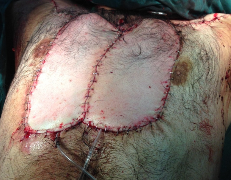
Figure 4. Reconstruction with deltopectoral fasciocutaneous flaps based on the upper perforation of the internal mammary artery.
The authors harvested deltopectoral staged fasciocutaneous flaps bilaterally, based on the mammary artery perforators. The flaps were rotated to cover the defect, and the harvest site was primarily closed. The final pathology showed a CD34 positive tumor that was ASMA, S100, CD39, and BC12 negative. The immunohistochemical results favored sarcoma (a fibrosarcomatous tumor arising in dermatofibrosarcoma protuberans). All margins were negative with close deep margins. The final tumor stage was Stage I: primary tumor, localized disease.
References
- Madan RK, Ferzil GM, Gallitano SM, Chen C-SJ, Siegel DM. Dermatofibrosarcoma protuberans clinical presentation. Eds James WD, Wells, MJ, Albertini JG. http://emedicine.medscape.com/article/1100203-clinical#b5. Medscape. Updated August 16, 2018. Accessed December 10, 2018.
- Lemm D, Mugge LO, Mentzel T, Hoffken K. Current treatment options in dermatofibrosarcoma protuberans. J Cancer Res Clin Oncol. 2009;135(5):653-665.
- Ugurel S, Kortmann RD, Mohr P, Mentzel T, Garbe C, Breuninger H. Short German guidelines: dermatofibrosarcoma protuberans. J Dtsch Dermatol Ges. 2008;6 Suppl 1:S17-S18.


