|
Illustrations
and Imaging |
page
35 |
Stage
IIIA
 |
 |
|
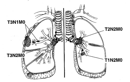
Fig.
42: Diagram,
stage IIIA. Stage IIIA
disease includes tumors
with localized, circumscribed
extrapulmonary extension
and ipsilateral intrapulmonary
(including hilar) lymph
node metastasis, the
T3 N1 M0 subset, and
T1, T2, and T3 tumors
with metastasis limited
to the ipsilateral mediastinal
and subcarinal lymph
nodes, the T1 N2 M0,
T2 N2 M0, T3 N2 M0 subsets. |
|
|
 |
|
|
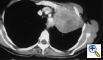
Fig.
43: Computed
tomographic scan of bronchogenic
carcinoma involving the
anterior chest wall.
The medial pleura was
not involved. Metastatic
disease was present in
a normal sized left hilar
lymph node, T3 N1 M0,
stage IIIA disease. |
|
|
|
|
|
|
|
|
|
|
|
|