Robotic-Assisted Thoracoscopic Resection of a Secretory Intrapericardial Paraganglioma [1]
This video is part of CTSNet’s 2025 Resident Video Competition. Watch all entries into the competition, including the winning videos. [3]
In this video, the authors demonstrate a robotic-assisted thoracoscopic resection of a secretory intrapericardial paraganglioma. The purpose of this video is to review preoperative planning with a multidisciplinary team, robotic port placement, and surgical technique.
A 42-year-old woman presented to the emergency room with nonspecific symptoms, including abdominal and chest pain, anxiety, and syncope. Significant findings on physical examination included hypertension and firm swelling of the neck bilaterally.
Imaging of the neck revealed contrast-enhancing masses at the carotid bifurcations bilaterally, consistent with carotid body tumors, the most common type of paraganglioma in the neck. A positron emission tomography-computed tomography (PET-CT) scan, obtained to assess for multifocal extra-adrenal paragangliomas, identified a mediastinal mass with intense uptake measuring 2.6 x 1.9 cm. On CT angiogram of the chest, the mass was well-circumscribed within the pericardium, abutting the right atrial appendage and ascending aorta without any involvement of the coronary vessels.
Serum and urine metanephrines were elevated and confirmed the diagnosis of extra-adrenal paragangliomas.
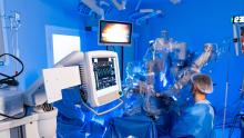
The patient consented to surgical resection. Preoperative planning included medical optimization (alpha blockade, beta blockade, and a high-salt diet) and engaging a multidisciplinary team. The surgical approach was planned via robot-assisted right video-assisted thoracoscopic surgery (VATS), with the possibility of converting to median sternotomy and even initiating cardiopulmonary bypass in the event of uncontrolled bleeding or hemodynamic compromise during resection. Therefore, a cardiac surgeon, cardiac anesthesiologist, and perfusion team were ready in the room.
After induction of general anesthesia with a double-lumen endotracheal tube, the patient was positioned supine with a bump under the right scapula. The patient's chest, abdomen, groin, and upper thighs were prepped and draped.
Four 8 mm robotic port sites were placed. The first was placed in the fifth intercostal space at the anterior midaxillary line. The chest was then insufflated with CO2 at a pressure of 8 mmHg. The remaining three ports were placed in third intercostal space in the axillary skin fold, the sixth intercostal space at the midaxillary line, and the seventh intercostal space at the midclavicular line.
Starting with a 0-degree endoscope, a long bipolar forceps in the right hand and a fenestrated bipolar forceps in the left hand, the mediastinal pleural was opened, and the thymus was teased away from the pericardium.
The pericardium was opened, revealing a 2 cm, extremely vascular tumor which appeared to be stuck to the anterior wall of the ascending aorta, away from the superior vena cava (SVC) and coronary arteries. The pericardium was tented up with a 2-0 silk suture to the sternum. A cigar sponge was used to gently retract the right atrial appendage caudad. No other lesions were seen.
Using a long bipolar initially, a plane was created between the mass and the adventitia of the aorta. The instruments were then switched to a vessel sealer, and the tumor was carefully dissected circumferentially off the aortic adventitia.
Hemostasis was achieved using the vessel sealer bipolar cautery, a hemostatic agent, and surgical adhesive. A 19-French Blake drain was placed in the pericardium. Final pathology was consistent with a mediastinal paraganglioma.
References
- Naser JA, van Zyl M, Gruber LM, Gulati R, Friedman PA, Young WF Jr, Hemmati P, Foley TA, Schaff HV, Crestanello JA, Pislaru SV. Role of Multimodality Imaging and Preoperative Management in Intrapericardial Paragangliomas: Experience From a Case Series. JACC Case Rep. 2022 Jul 20;4(14):871-877.
- Ojiaku M, Peña E, Belanger EC, Chan KL, Dennie C. Functioning intrapericardial paraganglioma: multimodality imaging findings and pathological correlation. Circulation. 2014 Oct 14;130(16):e137-9.
Disclaimer
The information and views presented on CTSNet.org represent the views of the authors and contributors of the material and not of CTSNet. Please review our full disclaimer page here. [4]