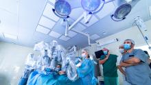
Robotic Left Lower Lobectomy With Bronchoplasty [1]
Introduction
The authors present the case of a 77-year-old male with a significant smoking history who underwent a robotic left lower lobectomy with bronchoplasty. Computed tomography (CT) imaging revealed a left lower lobe opacification and concern for malignancy, which was intensely fluorodeoxyglucose (FDG) avid on positron emission tomography (PET) scan. Bronchoscopy revealed an endobronchial mass in the distal left lower lobe bronchus. A biopsy confirmed squamous cell carcinoma, clinical stage T2bN0, with programmed cell death ligand 1 (PD-L1) expression of less than one percent. After multidisciplinary discussion, neoadjuvant treatment was initiated with four cycles of carboplatin, pembrolizumab, and paclitaxel. Restaging CT demonstrated a good response to treatment. Preoperative pulmonary function tests (PFTs) and exercise capacity were suitable for operative resection. The patient was then brought to the operating room for a left lower lobectomy with bronchoplasty to address the persistent mass in the left lower lobe bronchus.
Key Technical Steps
Key technical steps included adhesiolysis to mobilize the left upper and lower lobes, alongside mediastinal lymphadenectomy that involved stations 9L, 7L, 10L, 5L, and 6L. Dissection and identification of hilar structures were performed, including the lingular vein and artery, inferior pulmonary vein, and the upper and lower lobe bronchi, followed by parenchymal division. The basilar and superior segmental arteries and the inferior pulmonary vein were subsequently divided.
Bronchoplasty of the left lower lobe bronchus was conducted while retracting and preserving the left upper lobe bronchus and superior pulmonary vein. The posterior membranous wall of the bronchus was approximated to the cartilaginous wall with interrupted Vicryl sutures followed by a running polydioxanone (PDS) suture to ensure an airtight seal. A thymic flap was harvested and attached to reinforce the bronchoplasty seal.
The final pathology revealed invasive squamous cell carcinoma staged as pT1bN0M0, with negative surgical margins and no lymphovascular invasion or lymph node involvement. The patient's postoperative course was complicated by a prolonged air leak; however, he was discharged on postoperative day seven after resolution. Follow-up visits showed that the patient was doing well. The patient was seen by medical oncologists, who recommended an additional one-year course of pembrolizumab.
Conclusion
This case demonstrates the successful application of robotic left lower lobectomy with bronchoplasty in a patient with invasive squamous cell carcinoma. Bronchoplasty was necessary to ensure complete oncologic resection while preserving the upper lobe bronchus. This video underscores the value of minimally invasive techniques in complex airway reconstruction and serves as a technical reference for trainees and surgeons managing similar cases.
Disclaimer
The information and views presented on CTSNet.org represent the views of the authors and contributors of the material and not of CTSNet. Please review our full disclaimer page here. [3]