T4
 |
 |
|
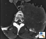
Fig.
18b: Computed
tomographic scan of the
chest at the 3rd thoracic
vertebra shows invasion
of the left side of the
vertebral body and extension
of tumor into the intervertebral foramen (white arrow), T4. |
|
|
 |
|
|
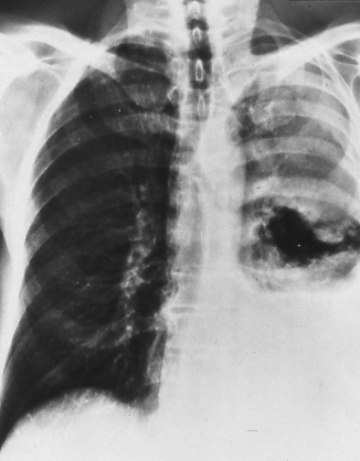
Fig.
19: Posterior-anterior
chest radiograph showing
pleural effusion on the
left. Malignant pleural
effusion due to the underlying
malignant process is
classified T4, whether
cytology is positive
or negative. |
|
|
A few patients are seen in whom cytopathological
examination of pleural fluid on more than
one specimen is negative for tumor, the
fluid is non-bloody and is not an exudate.
If negative cytologies and clinical judgment
indicate that the effusion is unrelated
to the lung cancer, the effusion is disregarded
as a staging element and T1, T2 or T3 as
appropriate is assigned.
|