The patient was a 67-year-old man who was referred for surgical revascularization of coronary artery disease. Three months earlier, he had undergone emergency percutaneous coronary intervention (PCI) to the right coronary artery. He had a history of paroxysmal atrial fibrillation. Coronary angiography demonstrated significant proximal left anterior descending artery stenosis. An off-pump totally endoscopic coronary artery bypass (TECAB) using the left internal thoracic artery (LITA) to the left anterior descending artery (LAD) with concomitant left atrial appendage closure was planned.
Port Placement
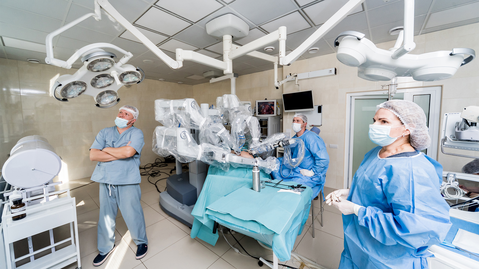
The patient was positioned supine with slight elevation of the left chest. An 8 mm camera port was placed in the fourth intercostal space at the anterior axillary line, and a 30-degree up camera was introduced. The right robotic arm port was inserted in the second intercostal space. The left robotic arm port was inserted in the sixth intercostal space. A 2 cm incision was made in the sixth intercostal space at the posterior axillary line, and a 12 mm port was inserted. The robotic system was docked from the patient’s right side.
Left Atrial Appendage Closure
The posterior pericardium was opened and extended toward the pulmonary artery to widely expose the left atrial appendage. The base of the appendage was measured, and a 40 mm Atriclip was selected and introduced through the port. The appendage was gently mobilized without injury while the clip was carefully advanced to the base. After confirming satisfactory positioning by transesophageal echocardiography (TEE), the clip was deployed by the bedside assistant.
Closed-Chest Stabilizer Technique
An anterior pericardiotomy was performed, and the left anterior descending artery was identified. The left internal thoracic artery was harvested in a skeletonized fashion. The shaft of a stabilizer was introduced through the subcostal incision. The port was temporarily removed, and the head of the stabilizer was inserted through the 2 cm incision. The port was then reinserted to maintain CO2 insufflation. Inside the CO2 pressurized chest, the stabilizer shaft and head were connected.
LITA-LAD Bypass
The stabilizer was positioned over the target LAD. The surface of the LAD was dissected using low-energy electrocautery at 10 watts. A silastic snare suture was placed around the LAD. Systemic heparin was administered to maintain an active clotting time (ACT) level greater than 300 seconds. The distal LITA was clipped. A small arteriotomy was created, and an epidural catheter was inserted for intraluminal papaverine injection. The length of the LITA was adjusted, and it was secured to the pre-made tie on the stabilizer at the appropriate length. The LAD was snared. A small coronary arteriotomy was made with the coronary knife and extended using Potts scissors. A coronary shunt was inserted, and the snare was released.
The LITA to LAD anastomosis was performed using 7-0 3 cm long suture. A transit-time flow measurement probe was introduced through the port. Graft flow measured 113 ml/min with a pulsatility index of 1.8.
Postoperative Course
The patient was extubated in the operating room. The postoperative course was uneventful, and he was discharged home on postoperative day one.
Disclaimer
The information and views presented on CTSNet.org represent the views of the authors and contributors of the material and not of CTSNet. Please review our full disclaimer page here.
 839 views on Vimeo
 1,744 views