A 63-year-old woman presented with myasthenia crisis and ptosis, for which she was treated with immunoglobulin. Her medical history included type 2 diabetes mellitus (DM), hypertension, and hypothyroidism.
A computed tomography (CT) scan of the chest showed a well-defined, isodense soft tissue lesion with homogenous post contrast enhancement (30 x 29mm) seen in the anterior mediastinum. The lesion was observed abutting the aorta and the main pulmonary artery (MPA) posteriorly. There was no evidence of fat attenuation, calcification, or hemorrhage within the lesion. An electrocardiogram (ECG), echocardiogram and coronary angiogram were normal. The patient was started on tablets of azathioprine and pyridostigmine.
She was planned for a radical thymectomy, and two doses of immunoglobulin were given before surgery to prevent perioperative myasthenia crises.
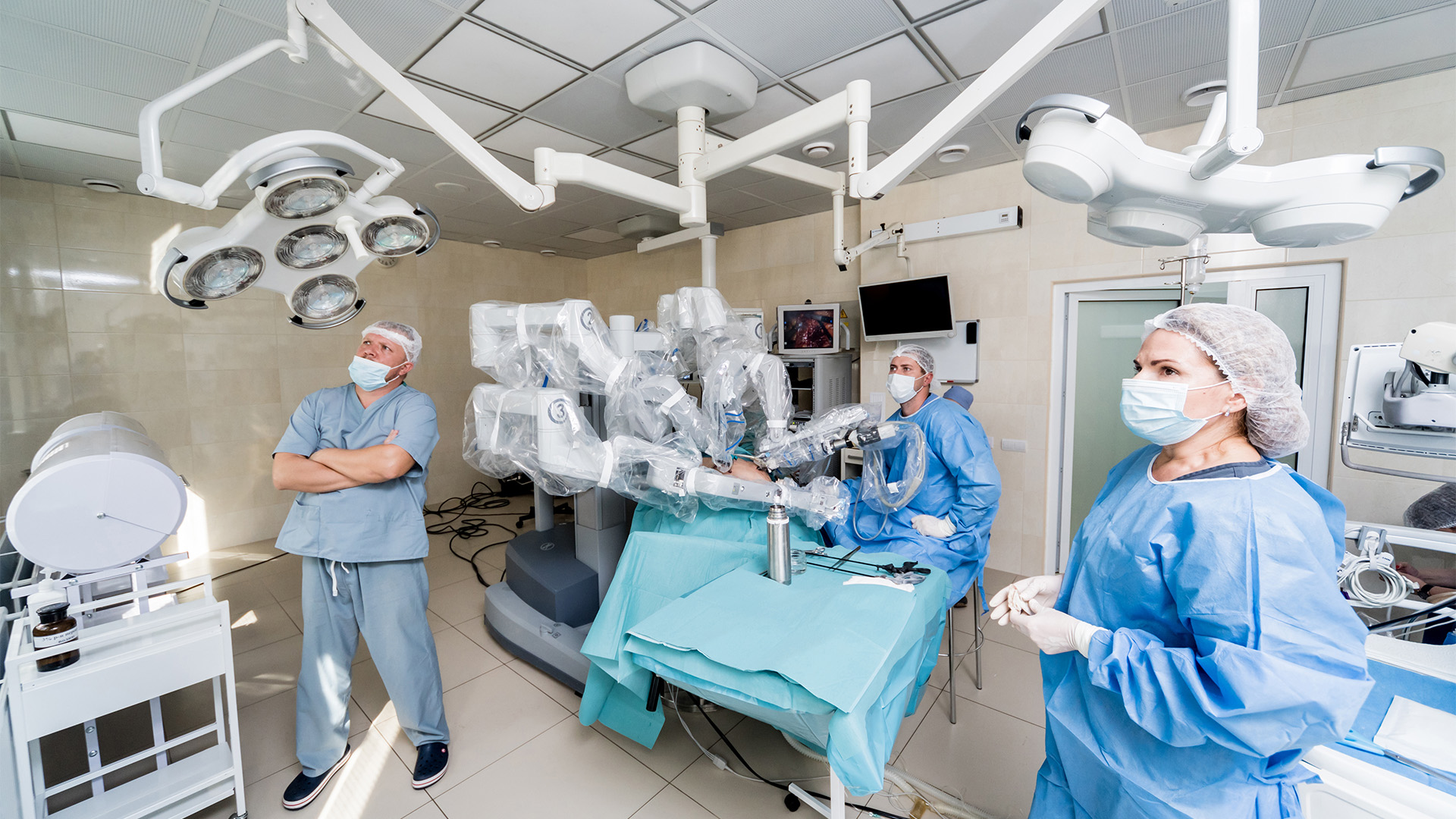
She underwent a robotic radical thymectomy using three ports from the left side, as the thymoma was located on the left side. This approach is ideal for masses growing mainly on the left side of the chest and allows for easy to reach ectopic (wandering) tissue near the left phrenic nerve and the aortopulmonary window.
Under general anesthesia with double-lumen intubation, the patient was positioned in a 30-degree semi-supine position, left side up, with a roll placed under the left shoulder for better left chest exposition. The right arm was right extended on a padded board in case right-sided ports were required.
Carbon dioxide (CO2) insufflation was started with a 6l/min flow and 8 mmHg pressure at the upper port site through Veress needle to prevent damage of mediastinal structures. The first incision (camera port) was performed in the fifth intercostal space at the anterior axillary line. The two operative ports were performed at the anterior axillary line in the third intercostal space and at the fifth intercostal space at the mid-clavicular line.
The thymic mass was removed along with mediastinal fat from both sides. Bilateral chest tubes were inserted. She was extubated on the table and continued taking tablets of azathioprine and pyridostigmine. Her ptosis was resolved, and she was discharged on the fifth postoperative day. Histopathological examination showed a type B2 thymoma, with no areas of thymic carcinoma noted. The tumor infiltrated into the mediastinal fat, but the lymph nodes were free of tumor (pT1aN0).
The left-sided robotic assisted extended thymectomy is a safe procedure that facilitates mediastinal mass resection. The working space is smaller because the heart is located on the left side, requiring an experienced surgical team.
References
- Raza B, Dhamija A, Abbas G, Toker A. Robotic thymectomy for myasthenia gravis surgical techniques and outcomes. J Thorac Dis. 2021;13(10):6187-6194.
- Ferrari-Light D, Cerfolio RJ. Left-sided approach for robotic thymectomy: technical tips, advantages and drawbacks. Shanghai Chest 2019:3:3.
Disclaimer
The information and views presented on CTSNet.org represent the views of the authors and contributors of the material and not of CTSNet. Please review our full disclaimer page here.
 202 views on Vimeo
 477 views