


Ana Pita Fernandez

Enlarging Axillary Incision The axillary incision was enlarged in two ways. First, a horizontal conversion to a posterolateral approach was performed based on previous pen markings. Additionally, a vertical downward enlargement was made to reach...

This article is part of CTSNet’s Guest Editor Series, Coronary Arterial Anomalies—Pediatric and Adult Congenital. CTSNet Senior Editor Dr. Sameh Said invited both pediatric and adult cardiac surgeons from around...

This case was performed during a humanitarian mission in Paraguay in April 2023. The patient is a ten-year-old, 40 kg girl with severe tricuspid regurgitation. The surgical team found annular...

Introduction Levy et al [1] first described the aorto-left ventricular tunnel in 1963. This rare congenital malformation is an abnormal channel that connects the ascending aorta with the left ventricle,...

Donation after Circulatory Death (DCD) is gaining worldwide acceptance as an adjunct to brain death (BD) in transplant surgery. Initially performed for abdominal organs procurement, DCD was then carried out...

Introduction Double aortic arch is a relatively rare cardiovascular malformation. It is an anomaly of the aortic arch in which two aortic arches form a complete vascular ring that can...
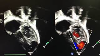
A 2-week-old, 3.5 kg child was transferred from a distant hospital (300 miles away) on congestive heart failure. Suspected diagnosis was ventricular septal defect (VSD), atrial septal defect (ASD), and...

IntroductionVascular ring is a congenital anomaly of the aortic arch that compresses the esophagus and/or trachea. It can be asymptomatic for life and not require any intervention. On the contrary,...

This video describes a simple method of complete reconstruction of a truncal valve using equine pericardium in a one-year-old patient with severe truncal valve regurgitation. Surgical Technique Through a transverse...
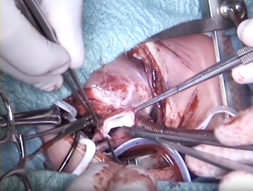
Introduction Hemitruncus is an extremely uncommon anomaly, accounting for 0.1% of all congenital heart diseases (1). This defect is defined by the anomalous origin of one pulmonary artery, most commonly...
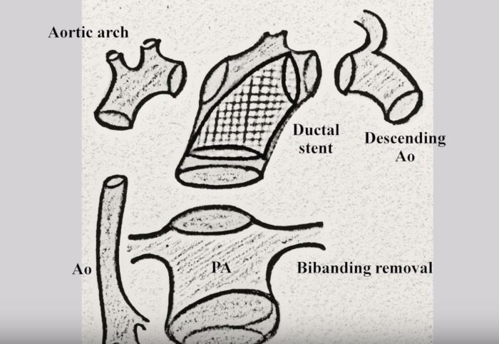
Technique A 3.2 kg male neonate was delivered with the diagnosis of aortic atresia, ventricular septal defect, balanced ventricles, hypoplastic ascending aorta (1.5 mm), and severe hypoplastic aortic arch. On...
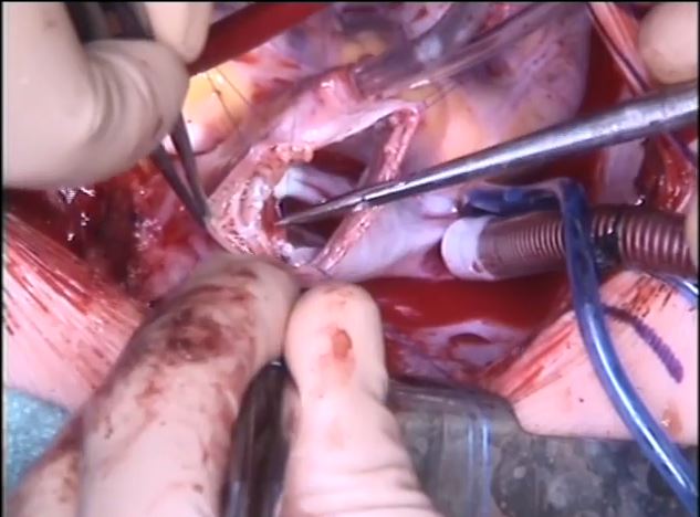
Partial anomalous pulmonary venous connections (PAPVC) are a very heterogeneous group of lesions, in which at least one pulmonary vein is draining into the systemic venous system. These congenital lesions...