


Congenital & Pediatric

This study demonstrates that using autologous right atrial appendage (RAA) tissue to create a neovalve during RVOT reconstruction is a safe and effective alternative for pediatric patients with congenital heart disease. The technique showed promising...

Diaphragmatic paralysis following congenital cardiac surgery is an important cause of postoperative respiratory morbidity in neonates and infants. Prompt recognition and appropriate management are essential to reduce ventilator dependence and...


 …
…This week on The Beat, CTSNet Editor-in-Chief Joel Dunning spoke with James Cox, Professor of Cardiac Surgery at the Feinberg School of Medicine at Northwestern Medicine in Chicago, Illinois, USA,...

An 8-year-old male (24 kg) presented with intermittent fever for 45 days. Echocardiography demonstrated infective endocarditis of a bicuspid aortic valve with multiple vegetations (largest 8 × 6 mm), severe...

This video demonstrates the modified trapdoor technique for direct reimplantation of an anomalous left coronary artery from the pulmonary artery. Case Presentation Following cardiopulmonary resuscitation, a three-month-old, 4.7 kg infant,...
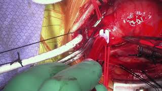 …
…A two-week-old, 3.5 kg newborn presented with tachypnea and a heart murmur. An echocardiogram showed a type I aortopulmonary window and patent foramen ovale. A computed tomography scan confirmed the...
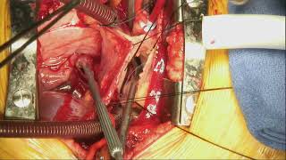
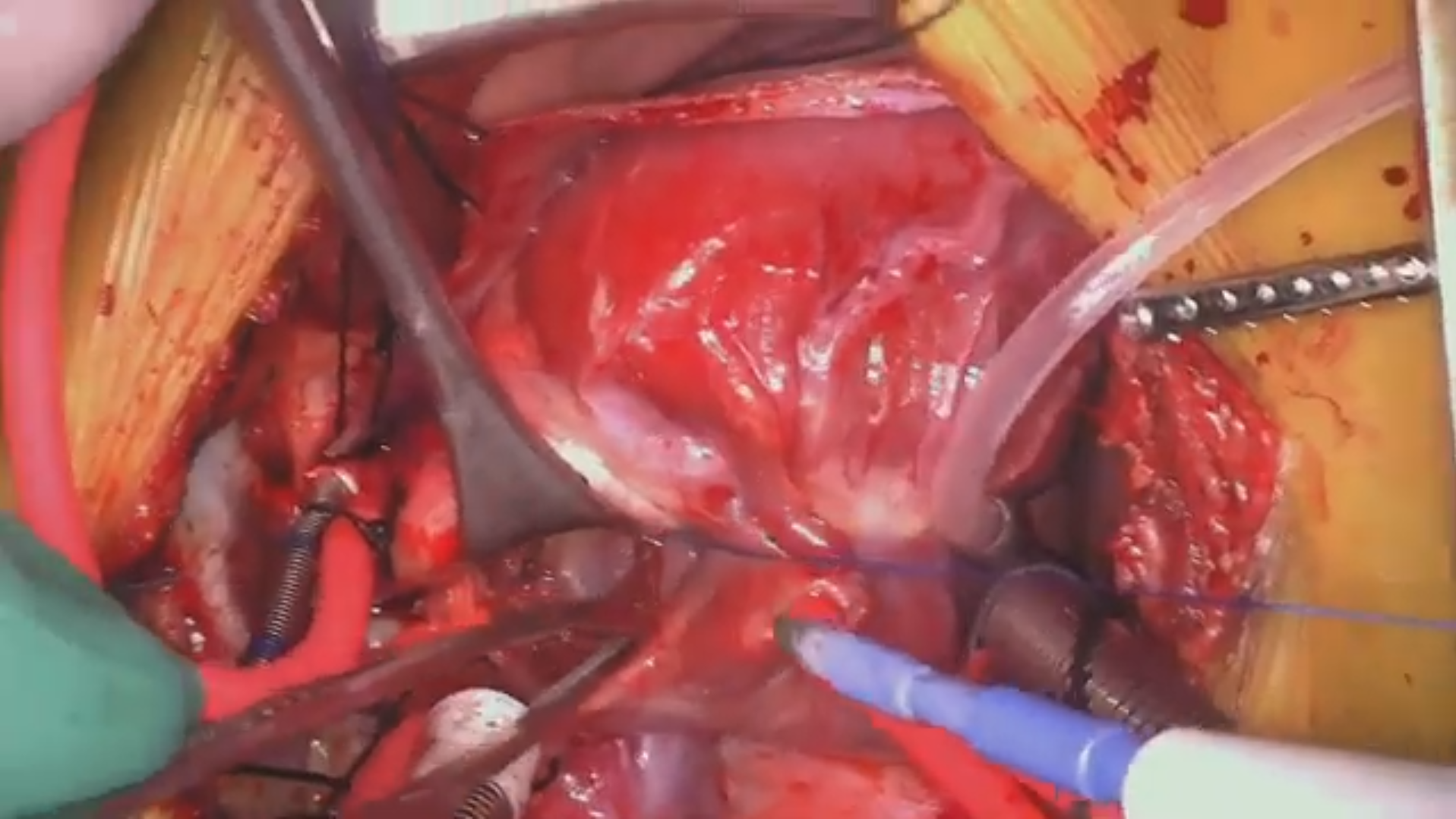
A sixty-two-year-old female with a history of chronic cough and exercise intolerance initially presented for elective calcium scoring via cardiac CT. The scan showed previously undiagnosed anomalous pulmonary venous return...
 …
…As this article and video demonstrate, the superior approach for repair of supracardiac TAPVC carries several advantages, including minimal-to-no heart manipulation, ease of accessibility through the transverse pericardial sinus, and...
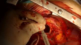
Mitral valve replacement using the top-hat technique can be an option in patients with small mitral annulus or patients with patient-prosthesis mismatch. This technique can provide adequate left ventricle in-flow...
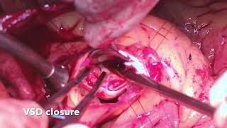
A thirty-year-old male was admitted for decompensated heart failure. He had a history of a hole in the heart during childhood. The echocardiography on admission showed left ventricular function, an...
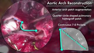
This video demonstrates Dr. James Tweddell’s surgical strategy in a two-month-old male with diagnosis of aortic atresia, hypoplastic aortic arch, coarctation, large perimembranous VSD, mildly hypoplastic mitral valve, normal biventricular...
 …
…This video presents a twenty-two-year-old male born with congenital aortic stenosis secondary to a bicuspid aortic valve who had two previous sternotomies. He received an initial open aortic valvotomy at...
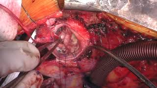
A two-month-old female presented with failure to thrive and shortness of breath during feedings. Echo images show ed type 2 aortopulmonary window, left ventricular enlargement, and systemic pulmonary hypertension. After...