ALERT!
This site is not optimized for Internet Explorer 8 (or older).
Please upgrade to a newer version of Internet Explorer or use an alternate browser such as Chrome or Firefox.
Intraoperative Angioscopy for Endoscopic Veins: A Simple Quality Control Measure
Background
Arterial grafts remain the conduit of choice for most coronary surgeons. However, saphenous vein grafts are the most commonly used conduits. Endoscopic vein harvest (EVH) has become the procedure of choice in harvesting saphenous veins for coronary artery bypass grafting (CABG). Studies have shown this technique to be a reliable alternative to the open technique [1,2]. The technique of EVH has been associated with intraluminal clot [3,4].
Our strategy addresses two of the three variables in Virchow’s triad which are thought to contribute to thrombosis: (1) alterations in blood flow (stasis), (2) vascular endothelial injury, and (3) alterations in the blood (hypercoagulability). Our two strategies address the coagulation and stagnation arms of Virchow’s triad: (1) pre-heparinization (improving coagulation status), and (2) open carbon dioxide (CO2) insufflation system for endoscopic vein harvesting or limiting the amount of CO2 pressure during sealed carbon dioxide EVH, therefore limiting stagnation of blood. Pre-heparinization is the most studied yet some feel that there are drawbacks as many surgeons are concerned with the risk of bleeding. Despite pre-heparinization, 11% of veins harvested may still have intraluminal clot [4]. A quality control measure is needed to identify those patients who have clot. Directed therapy such as Fogarty thrombectomy or thrombolysis can then be performed on those vein segments.
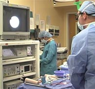
Optical coherence tomography (OCT) is an intraluminal imaging technique that is expensive and experimental and not widely available in most operating rooms. Intraoperative angioscopy was used by vascular surgeons in the early 90’s but abandoned with the advent of endovascular techniques. We describe a technique for intraoperative vein angioscopy for EVH, a technique that evaluates the entire length of conduit intraoperatively before grafting, and usually adds only three minutes to the operative procedure.
Material and Methods
Following a median sternotomy, the patient is pre-heparinized with 5000 units of intravenous heparin, followed by incremental heparin doses to bring the activated clotting time above 300 seconds. Administration of Amicar® is delayed until the EVH is completed. All patients undergo concurrent left internal thoracic artery harvest and EVH with EVH being performed using the VasoView® 7 system (MAQUET Cardiovascular LLC, San Jose, CA). A conical dissection tip is attached to the endoscope in order to make a dissection tunnel around the greater saphenous vein. A seal is created around the tunnel using trochar port balloon. CO2 is instilled to a pressure of 10-12 mmHg, but not to exceed the patient’s central venous pressure. Division of branches is preformed with bipolar electrocautery (35 watts). Proximal and distal vein ligation is accomplished through separate stab incisions.
Following its removal, the vein is carefully prepared and gently flushed with isotonic saline. A vessel cannula (Sorin® coronary cardioplegia adaptor, Sorin Group USA, Arvada CO) is then attached to the distal end of the vein, followed by external visual inspection of the vein. Once the vein is satisfactory, an atraumatic clamp (Fogarty® soft jaw 6mm clamp model 614-06, Baxter-Edwards, Santa Ana CA) is then placed on the proximal end. Taking care not to over-distend the vein, it is then filled with an isotonic saline solution. The 1.4mm ultrathin Olympus® fiberoptic angioscope (Olympus America, Inc., Center Valley PA) is introduced through the vessel cannula and into the vein. The angioscope is guided to the end of the vein. No attempt is made to inspect the vessel until the withdrawal of the angioscope at which time the inspection process is begun.
Normally, the endoluminal aspect of the vein is smooth and glistening with natural corrugations as well as valves. Intraluminal clots can vary in size with clots tending to form around points of stagnation such as a valves or the entrance of tributary branches.
Comment
EVH has become the procedure of choice at many cardiac surgery centers. Approximately 80% of cardiac surgery centers offer EVH as a modality for saphenous vein harvest. Randomized trials have shown the benefits of veins harvested with this less invasive modality, including a lower infection rate, fewer wound complications, improved cosmesis and greater patient satisfaction and histological studies have revealed no significant difference with the endothelial integrity [1, 2]. Intraluminal clot after EVH has been previously described [3, 4]. There are few techniques available to rapidly and effectively image conduits for CABG intraoperatively prior to grafting, so the extent and distribution of intraluminal clot following EVH has been difficult to evaluate. Recently, the Prevent IV multicenter trial showed a low short-term vein graft patency rate associated with EVH. The odds ratio was 1.35 (p< 0.001) [5]. In addition, a study by Posson, et al, showed that intraluminal clot occurred in up to 80% of veins harvested by the EVH [4].
We feel the use of a flexible angioscope to image the endoluminal surface of harvested saphenous veins provides a rapid intraoperative assessment of the entire length of vein conduit prior to grafting in CABG patients. This modality provides a quality control measure in evaluating saphenous veins, where further intervention could be implemented if warranted or improved prevention of intraluminal clot could be devised.
The video can also be viewed, larger, in a new window: Intraoperative Angioscopy for Endoscopic Veins: A Simple Quality Control Measure.
References
- Kiaii B, Moon BC, Massel D, et al. A prospective randomized trial of endoscopic versus conventional harvesting of the saphenous vein in coronary artery bypass surgery. J Thorac Cardiovasc Surg 2002;123:204-12.
- Puskas JD, Wright CE, Miller PK, et al. A randomized trial of endoscopic versus open saphenous vein harvest in coronary bypass surgery. Ann Thorac Surg 1999;68:1509-12.
- Burris N, Schwartz JB, Kwon M, Poston R. Incidence of residual clot strands in saphenous vein grafts after endoscopic harvest. Innovations 2006;1:323-27.
- Brown EN, Kon ZN, Tran R, et al. Strategies to reduce intraluminal clot formation in endoscopically harvested saphenous veins. J Thorac Cardiovasc Surg 2007;134:1259-65.
- Alexander JH, Hafley G, Harrington RA, et al. Efficacy and safety of edifoligide, an E2F transcription factor decoy, for prevention of vein graft failure following coronary artery bypass graft surgery: PREVENT IV-a randomized controlled trial. JAMA 2005:294:2446-54.
- Burris N, Schwartz K, Tang C-M, et al. Catheter-based infrared light scanner as a tool to assess conduit quality in coronary artery bypass surgery. J Thorac Cardiovasc Surg 2007;133:419-27.


