Case Presentation
A 55-year-old female was evaluated for a large, asymptomatic Morgagni hernia containing the transverse colon and a significant portion of the greater omentum that had been incidentally noted on imaging. Preoperative imaging demonstrated a sizable anterior diaphragmatic defect measuring approximately 30 cm². Elective operative repair of this hernia was chosen. Given the size of the defect and hernia contents, a robotic transabdominal preperitoneal approach was selected to allow optimal visualization, safe reduction, and durable mesh reinforcement.
The Surgery
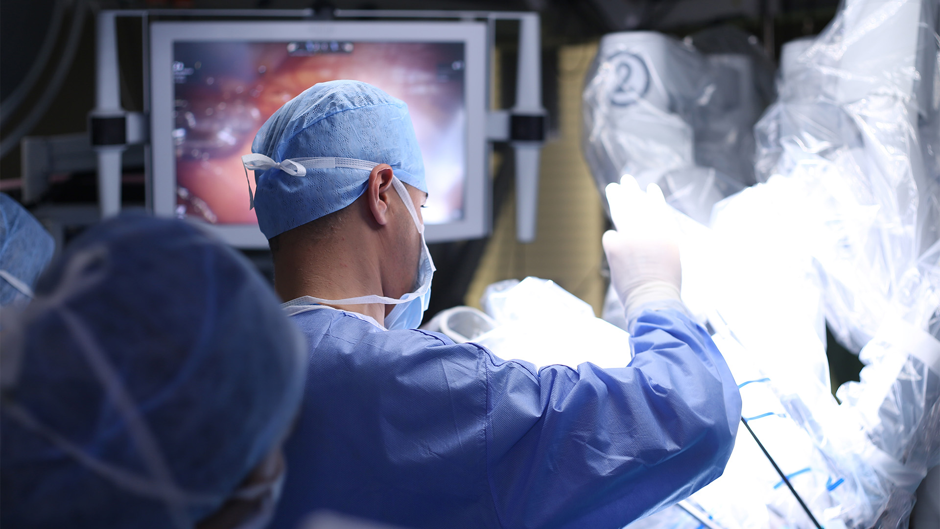
The operation was performed using a robotic platform with standard foregut positioning.
The procedure is demonstrated in nine key steps:
Step 1: Patient Positioning and Robotic Setup
The patient was positioned in standard foregut configuration, and four 8 mm robotic ports were placed to allow optimal access to the anterior diaphragm and mediastinum.
Step 2: Initial Exploration
A diagnostic survey of the abdomen confirmed the presence of a large Morgagni hernia and defined the extent of the defect and hernia contents.
Step 3: Reduction of Hernia Contents
The transverse colon and the majority of the greater omentum were carefully reduced from the thoracic cavity into the abdomen using an atraumatic technique.
Step 4: Development of the Preperitoneal Plane
Attention was turned to the diaphragmatic defect, and a preperitoneal plane was developed to facilitate a transabdominal preperitoneal repair.
Step 5: Creation of the Peritoneal Flap
A peritoneal flap was created and mobilized posterior to the diaphragm, providing coverage for subsequent mesh placement.
Step 6: Assessment of the Defect
The diaphragmatic defect was fully exposed and assessed, confirming its suitability for primary closure with mesh reinforcement given the large defect size.
Step 7: Primary Closure of the Defect
The defect was closed primarily using absorbable barbed sutures, restoring diaphragmatic continuity while minimizing tension.
Step 8: Mesh Placement
Polypropylene mesh was placed in the preperitoneal space to reinforce the repair and provide durable coverage of the approximately 30 cm² defect.
Step 9: Closure of the Peritoneal Flap and Final Inspection
The peritoneal flap was closed over the mesh with sutures, isolating the prosthesis from intra-abdominal contents. The repair was inspected to confirm adequate coverage, hemostasis, and proper diaphragmatic contour.
Postoperative Course
The patient tolerated the procedure well with an uncomplicated postoperative recovery. At follow-up, no postoperative symptomatology was reported, and the repair remained clinically intact.
This case highlighted that a robotic transabdominal preperitoneal approach allows excellent visualization and controlled mesh placement for large Morgagni hernia defects.
References
- Kaiser L. Mastery of Cardiothoracic Surgery, third edition 2014.
- Shields’ General Thoracic Surgery, eighth edition
Disclaimer
The information and views presented on CTSNet.org represent the views of the authors and contributors of the material and not of CTSNet. Please review our full disclaimer page here.
 182 views on Vimeo