


Introduction
Video-assisted thoracoscopic thymectomy (VATS) is an established approach for myasthenia gravis. The authors present a bilateral VATS thymectomy, highlighting key technical aspects.
Case Description
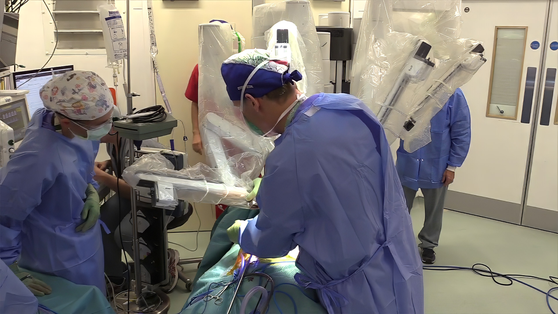
A 29-year-old male with ptosis was diagnosed with myasthenia gravis. Positron emission tomography (PET) imaging showed a 2 × 2 cm fluorodeoxyglucose (FDG)-avid anterior mediastinal lesion (standardized uptake value [SUV] 4.69). After medical optimization, bilateral VATS thymectomy was performed, starting from the left side to facilitate dissection and avoid early tumor handling. Complete en bloc resection of the thymus with mediastinal fibrofatty tissue, including both horns and epiphrenic fat, was achieved. Careful dissection along the innominate vein was performed while preserving the phrenic nerves and ensuring complete left horn delivery.
Results
Recovery was uneventful, and histopathology showed encapsulated Type B2 thymoma (Masaoka Stage I).
Disclaimer
The information and views presented on CTSNet.org represent the views of the authors and contributors of the material and not of CTSNet. Please review our full disclaimer page here.
 223 views on Vimeo







2 Comments
Dr. Mor, congratulations on your excellent video. Curious, how the audio was created for this video? Thank you for sharing your work with us.
Thank you so much for your kind words and encouragement.
For the narration, we used text-to-speech software during post-production editing to create clear and synchronized audio commentary for the operative steps.
Thank you for watching and for your support.