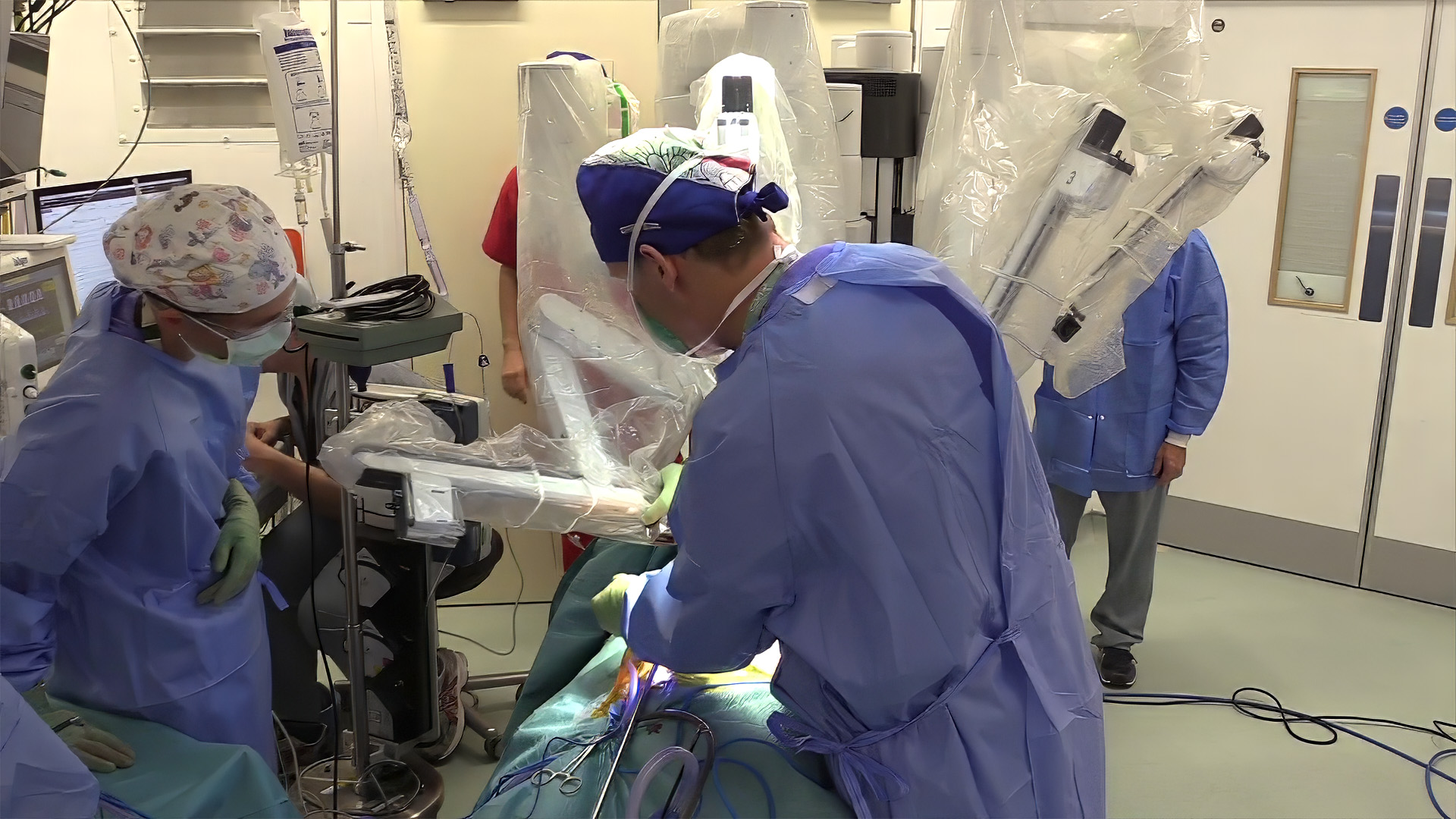
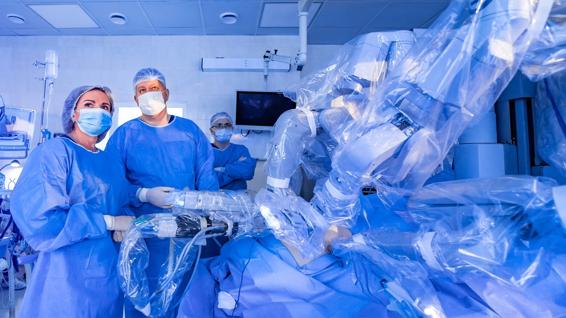
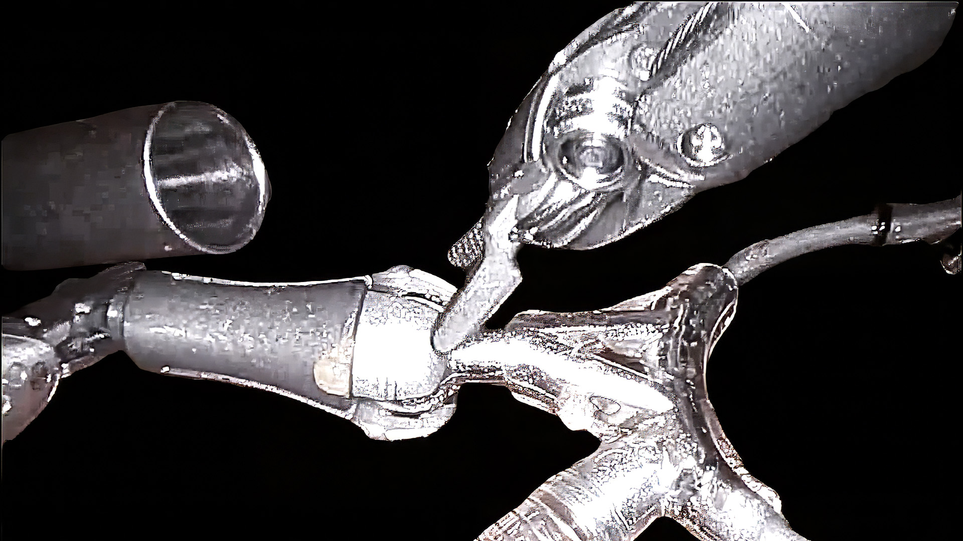
This video demonstrates the modified trapdoor technique for direct reimplantation of an anomalous left coronary artery from the pulmonary artery.
Case Presentation
Following cardiopulmonary resuscitation, a three-month-old, 4.7 kg infant, was referred to the authors’ institution with symptoms of heart failure and was diagnosed with an anomalous origin of the left main coronary artery from the pulmonary artery (ALCAPA).
An echocardiogram revealed significant left ventricular dysfunction with a dilated left ventricle and moderate mitral valve regurgitation. The diagnosis was also confirmed by computerized angiography, which showed the origin of the ALCAPA from sinus 1. The decision was made to proceed with surgical repair using a modified trapdoor approach for coronary transfer.
The Surgery
After a standard median sternotomy, the pericardium was opened, and a patch was harvested for subsequent use. The pulmonary artery branches were dissected and secured with silk snares. A standard cardiopulmonary bypass was initiated with cannulation of the aorta and the superior and inferior vena cava, and the aorta was cross-clamped.
Del Nido cardioplegia was administered simultaneously into the aortic and pulmonary roots. The tourniquets on both branch pulmonary arteries were snared to prevent run-off cardioplegia.
The pulmonary trunk was then transected, and the origin of the ALCAPA was identified and probed.
A large coronary button containing the whole pulmonary sinus tissue was excised, and the proximal left coronary artery (LCA) was extensively mobilized. A transverse aortotomy into the left coronary sinus was performed, and at the base of the sinus of Valsalva, the incision was reversed, creating a medially hinged trapdoor. The mobilized LCA was then directly reimplanted using the trapdoor technique to minimize tension on the coronary button. The aortic incision was closed with the same running suture, and a generous autologous pericardial patch was used to reconstruct the posterior sinus of the pulmonary artery.
After the aortic cross-clamp was removed, the main pulmonary artery was anastomosed in an end-to-end fashion during rewarming. The heart was weaned off bypass without difficulty. Transesophageal echocardiography showed a good flow into the LCA and improved left ventricular function.
The cardiopulmonary bypass and aortic cross-clamp times were 68 and 39 minutes, respectively. The patient was extubated on the fourth postoperative day and was discharged to a referral hospital on postoperative day 16 on ACEI therapy.
Left ventricular function continued to improve in the postoperative period and during follow-up. The last follow-up echocardiogram showed normal left ventricular function and size with no mitral valve regurgitation. The patient is doing well 12 years postoperatively.
Unlike the classic trapdoor technique, which utilizes only a limited portion of the left Valsalva sinus tissue, this modified approach involves a transverse aortotomy extending to the left Valsalva sinus, allowing full utilization of the sinus tissue in constructing the trapdoor.
The resulting larger trapdoor, combined with the incorporation of the entire pulmonary sinus tissue, facilitates a more anatomical orientation for coronary transfer and effectively reduces tension on the reimplanted coronary artery. In addition, this modification provides improved visualization of the reimplanted ALCAPA, the entire left Valsalva sinus, and the aortic valve.
References
- Said SM, Dearani JA, Burkhart HM, Schaff H V. Surgical management of congenital coronary arterial anomalies in adults. Cardiol Young. 2010;20 Suppl 3:68-85.
- Azakie A. Repair of Anomalous Coronary Artery From the Pulmonary Artery by Aortic Implantation. Operative Techniques in Thoracic and Cardiovascular Surgery. 2015;20(2):135-147.
- Sabateen F, Nosáľ M, Valentík P, Bobík L, Soják V. Anomalous Left Coronary Artery From the Pulmonary Artery With Ventricular Septal Defect and Double Aortic Arch: A Unique Case Report. World J Pediatr Congenit Heart Surg. 2024;15(4):508-510.
Disclaimer
The information and views presented on CTSNet.org represent the views of the authors and contributors of the material and not of CTSNet. Please review our full disclaimer page here.