Operative Steps
![[Illustration: Figure 1]](https://www.ctsnet.org/sites/default/files/graphics/experts/Adult/l_cohn_mininvas_mvr/4298_01.jpg) The patient is positioned supine, anesthetized and intubated with a single lumen endotracheal tube and then prepped and draped in the usual fashion. Defibrillator patches are placed on the patient’s back and anterior left chest wall. A transesophageal echo probe is placed to assess the pathology of the mitral valve disease and assist in air removal from the heart and document competency of the mitral valve repair at the completion of the procedure. A 6-8 cm skin incision is made at the lower end of the sternum beginning just below the xiphoid process.
The patient is positioned supine, anesthetized and intubated with a single lumen endotracheal tube and then prepped and draped in the usual fashion. Defibrillator patches are placed on the patient’s back and anterior left chest wall. A transesophageal echo probe is placed to assess the pathology of the mitral valve disease and assist in air removal from the heart and document competency of the mitral valve repair at the completion of the procedure. A 6-8 cm skin incision is made at the lower end of the sternum beginning just below the xiphoid process.
![[Illustration: Figure 2]](https://www.ctsnet.org/sites/default/files/graphics/experts/Adult/l_cohn_mininvas_mvr/4298_02.jpg) The incision is carried down to the sternum using cautery and the sternum is opened from below upward to the second intercostal space and extended into the interspace on the right. Care is taken to prevent injury to the right internal mammary artery and veins. The sternal retractor is the Koros® sliding CAB retractor.
The incision is carried down to the sternum using cautery and the sternum is opened from below upward to the second intercostal space and extended into the interspace on the right. Care is taken to prevent injury to the right internal mammary artery and veins. The sternal retractor is the Koros® sliding CAB retractor.
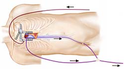
The cannulation technique is as follows: The ascending aorta is cannulated with a Sarns® 20 FR flexible cannula. ![[Illustration: Figure 3]](https://www.ctsnet.org/sites/default/files/graphics/experts/Adult/l_cohn_mininvas_mvr/4298_03.jpg) The superior vena cava is cannulated with a Baxter RMI® 24 FR right angle cannula and the inferior vena cava is cannulated from below by a Medtronic® percutaneous trans-femoral 21 French cannula extending to the mouth of the inferior vena cava with TEE guidance.
The superior vena cava is cannulated with a Baxter RMI® 24 FR right angle cannula and the inferior vena cava is cannulated from below by a Medtronic® percutaneous trans-femoral 21 French cannula extending to the mouth of the inferior vena cava with TEE guidance. ![[Illustration: Figure 4]](https://www.ctsnet.org/sites/default/files/graphics/experts/Adult/l_cohn_mininvas_mvr/4298_04.jpg) These small cannula are more than satisfactory for drainage of the venous return since assisted venous suction is used.
These small cannula are more than satisfactory for drainage of the venous return since assisted venous suction is used.
After cardiopulmonary bypass is established, the patient is cooled systemically to 28° C. The aorta is cross-clamped with an atraumatic clamp and cardiac arrest is achieved with cold blood cardioplegia in the![[Illustration: Figure 5a]](https://www.ctsnet.org/sites/default/files/graphics/experts/Adult/l_cohn_mininvas_mvr/4298_05a.jpg) ascending aorta. Since the patients do not have coronary artery disease, it is not mandatory to use retrograde cardioplegia. The right atrium is isolated by caval tourniquets and opened, and marsupialized.
ascending aorta. Since the patients do not have coronary artery disease, it is not mandatory to use retrograde cardioplegia. The right atrium is isolated by caval tourniquets and opened, and marsupialized.
The fossa ovalis is exposed and incised.
The left heart is decompressed. ![[Illustration: Figure 5b]](https://www.ctsnet.org/sites/default/files/graphics/experts/Adult/l_cohn_mininvas_mvr/4298_05b.jpg) The septal incision is carried cephalad about 2cm, but not so as to endanger the sinus node artery. Heavy silk marsupializing traction sutures are placed on the right and left edges of the interatrial septum and the left atrium exposed by retraction.
The septal incision is carried cephalad about 2cm, but not so as to endanger the sinus node artery. Heavy silk marsupializing traction sutures are placed on the right and left edges of the interatrial septum and the left atrium exposed by retraction.
Once the septum is opened and the retraction system has been set, anterior traction on the atrial septum is accomplished by a blade retractor from the Cosgrove retractor system to expose the mitral valve. To facilitate the exposure, coronary suckers or hand held flexible retractors can be used to move the left atrium to the right. Placing annuloplasty sutures in the posterior annulus and pulling these rightward may also help to expose the posterior annulus.
Mitral valve repair is then carried out in the usual fashion. In the case shown, P2 has ruptured chordae and is resected. Leaflet advancement is carried out with a running 4-0 Prolene® and then attachment of the two parts of the leaflet are put together with another running 4-0 Prolene® as well.
![[Illustration: Figure 6]](https://www.ctsnet.org/sites/default/files/graphics/experts/Adult/l_cohn_mininvas_mvr/4298_06.jpg)
![[Illustration: Figure 7]](https://www.ctsnet.org/sites/default/files/graphics/experts/Adult/l_cohn_mininvas_mvr/4298_07.jpg) The height of the leaflet after reconstruction is about 1-2 cms. Following completion of the valve resection repair a ring annuloplasty is done using a Cosgrove-Edwards® annuloplasty ring.
The height of the leaflet after reconstruction is about 1-2 cms. Following completion of the valve resection repair a ring annuloplasty is done using a Cosgrove-Edwards® annuloplasty ring.
Interrupted 2-0 multifilament mattress sutures beginning at the fibrous trigone and proceeding around the posterior annulus are placed through the ring. The handle is released and the annuloplasty ring slid into position. The sutures are tied and cut and the stent is removed leaving the annuloplasty ring in place. ![[Illustration: Figure 8]](https://www.ctsnet.org/sites/default/files/graphics/experts/Adult/l_cohn_mininvas_mvr/4298_08.jpg) The competency of the valve is then tested by injection of volumes of saline into the left ventricle, which also helps to displace air on the left atrium.
The competency of the valve is then tested by injection of volumes of saline into the left ventricle, which also helps to displace air on the left atrium.
The septal incision in the left atrium is closed using a continuous running 4-0 Prolene. Prior to the tying the last suture, air is removed by inflating the lungs. The aortic cross clamp is then removed and any residual air is removed by inflating the lungs and filling the left ventricle with blood and gentle suction through the cardioplegia site in the ascending aorta.
A pacing wire is placed on the anterior right ventricle prior to removal of the cross-clamp while the heart is decompressed so as to have good visualization of its placement.
After the cardiac function has been restored and the pacemaker wires brought out through the skin, two chest tubes are placed in the right chest and one in the pericardial cavity. The wound is closed in layers with stainless steel wire for the sternum, then layers of absorbable suture for the subcutaneous and fascia layers.
Preference Card
- DEBAKEY ILIAC CLAMPS
- CANNULAS
- COSGROVE EDWARDS ANNULOPLASTY RING
- KOROS SLIDING CAB RETRACT
Tips & Pitfalls
Safe cannulation will depend on the space available in the chest. In the vast majority of cases intrathoracic cannulation will be possible. If the femoral artery/femoral vein approach is chosen; the technique has been modified using 5cm supra inguinal ligament incision parallel to the inguinal ligament which has minimal wound complications. When the femoral artery/femoral vein is exposed purse string sutures are placed and the Seldinger technique is used to insert the cannulae rather than clamping and incising the vessels.
Placement of the annuloplasty sutures before repairing the valve helps by pulling the valve into the incision, increasing exposure.
References
Cohn LH. Mitral valve repair. Oper Tech Card Thorac Surg. 1998;3:109-25.
Aklog L, Adams DH, Couper GS, Gobezie R, Sears S, Cohn LH. Techniques and results of direct-access minimally invasive mitral valve surgery: A paradigm for the future. J Thorac Cardiovasc Surg 1998;116:705-15.
Byrne JG, Mitchell ME, Adams DH, Couper GS, Aranki SF, Cohn LH. Minimally invasive direct access mitral valve surgery. Seminars in Thoracic and Cardiovascular Surgery Vol 11, No 3 (July), 1999:212-22.
Cosgrove D, Aricidi J, Rodriguez et al: Initial experience with the Cosgrove-Edwards annuloplasty system. Ann Thorac Surg 60:499-504, 1995.