This video is part of CTSNet’s 2025 Resident Video Competition. Watch all entries into the competition, including the winning videos.
Clinical Presentation and Preoperative Course
The patient was a 56-year-old male with a history of hyperlipidemia, nephrolithiasis, and a recent episode of upper respiratory infection treated with antibiotics who presented with two months of persistent dyspnea. The patient had previously been a very active individual. Cross-sectional imaging done at an outside hospital demonstrated an elevated right hemidiaphragm but without other remarkable findings. Initial diaphragm fluoroscopy was concerning for right hemidiaphragm paralysis, with paradoxical motion on deep inspiration and during the sniff test.
Patient was referred to and evaluated in the authors’ thoracic surgery clinic and was eager to pursue surgical correction. The patient was presented with the options of traditional diaphragm plication and diaphragm pacing wire implantation. The patient was counseled extensively, making it clear that pacing wire implantation would only be feasible if there was some inherent phrenic nerve function upon exploration of the right hemidiaphragm. If there was no diaphragmatic excursion at all during exploration, then diaphragm plication would be done in the same operation. The patient decided to proceed with diaphragm pacing wire implantation first and understood the possibility of needing to convert to diaphragm plication.
Robotic-Assisted Laparoscopic Diaphragm Pacing Wire Implantation
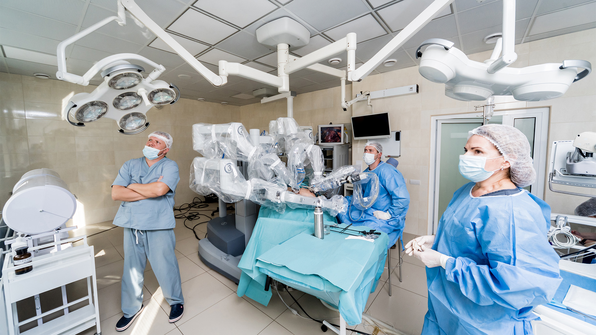
The abdomen was entered through direct vision using a trocar inserted through a 5 mm left upper quadrant incision. Two 8 mm robotic ports were then placed in the midline, approximately 12 cm inferior to the xiphoid, and in the right upper quadrant. The 5 mm left upper quadrant port was then exchanged for an 8 mm robotic port. Instruments used included the Cadiere forceps, a suture-cut needle driver, a vessel sealer, and the permanent cautery spatula.
After taking down the falciform ligament, the left and right hemidiaphragms were explored. The branches of the right phrenic nerve were stimulated using a cautery spatula connected to the pacing electrode externally. As expected, the right hemidiaphragm demonstrated weak but visible excursion. The left hemidiaphragm was then stimulated, demonstrating strong excursion. A decision was made at this time to proceed with the pacing wire implantation. Four internal pacing wire electrodes were then each tied with a 3-0 suture on the back table before being introduced into the abdomen for suturing. Two electrodes were placed in each hemidiaphragm at the appropriate locations along the phrenic nerve branches. The four electrodes were then brought out using the Carter-Thomason device. A grounding wire was then tunneled into the subcutaneous tissue. The wires were then coupled and placed in the cable adaptor device.
Postoperative Course
The patient tolerated the procedure well and was discharged on postoperative day one. At three weeks post-operation, the patient was tolerating diaphragm pacing at maximum settings without issues.
Key Takeaways
Robotic-assisted laparoscopic diaphragm pacing wire implantation is a novel, feasible, and reversible, surgical option for diaphragm paresis/paralysis with the potential to allow patients to regain inherent phrenic nerve and diaphragm function, given that there is some remaining phrenic nerve function upon exploration intraoperatively. Diaphragm plication can always be done as a second operation if patient does not show symptomatic improvement with pacing wires.
Disclaimer
The information and views presented on CTSNet.org represent the views of the authors and contributors of the material and not of CTSNet. Please review our full disclaimer page here.
 145 views on Vimeo
 574 views