Skip to content
Skip to content

The 6th Annual Innovation Video Competition Is Now Accepting Entries!

READ MORE
Not a member yet? Register now.
  • ABOUT
    • Leadership
    • Participation
    • Featured Sponsors
    • Submission Guidelines
    • Competitions
      • Innovation Video Competition
    • Community
      • Volunteer
      • Badges
    • Contact Us
  • EXPLORE CTSNET
    • CARDIAC
    • THORACIC
    • CONGENITAL
    • Clinical Care Professionals
    • JANS
    • Events
    • Journals
  • Videos
    • Atlas of Cardiac Surgery
    • Webinars
    • Master Class
    • Guest Interviews
    • Conference Sessions
  • Podcasts
    • The Beat With Joel Dunning
    • The Atrium
    • The Cardiac Recovery Room
    • The Lifeline
  • Profiles
    • Users Directory
    • Organizations
  • CAREERS
    • US Career Center
    • European Career Center
  • ABOUT
    • Leadership
    • Participation
    • Featured Sponsors
    • Submission Guidelines
    • Competitions
      • Innovation Video Competition
    • Community
      • Volunteer
      • Badges
    • Contact Us
  • EXPLORE CTSNET
    • CARDIAC
    • THORACIC
    • CONGENITAL
    • Clinical Care Professionals
    • JANS
    • Events
    • Journals
  • Videos
    • Atlas of Cardiac Surgery
    • Webinars
    • Master Class
    • Guest Interviews
    • Conference Sessions
  • Podcasts
    • The Beat With Joel Dunning
    • The Atrium
    • The Cardiac Recovery Room
    • The Lifeline
  • Profiles
    • Users Directory
    • Organizations
  • CAREERS
    • US Career Center
    • European Career Center
SUBMIT CONTENT
DONATE
JOIN

welcomes you to the new, improved website!

READ MORE
  • ABOUT
    • Leadership
    • Participation
    • Featured Sponsors
    • Submission Guidelines
    • Competitions
      • Innovation Video Competition
    • Community
      • Volunteer
      • Badges
    • Contact Us
  • EXPLORE CTSNET
    • CARDIAC
    • THORACIC
    • CONGENITAL
    • Clinical Care Professionals
    • JANS
    • Events
    • Journals
  • Videos
    • Atlas of Cardiac Surgery
    • Webinars
    • Master Class
    • Guest Interviews
    • Conference Sessions
  • Podcasts
    • The Beat With Joel Dunning
    • The Atrium
    • The Cardiac Recovery Room
    • The Lifeline
  • Profiles
    • Users Directory
    • Organizations
  • CAREERS
    • US Career Center
    • European Career Center
Not a member yet? Register now.
  • ABOUT
    • Leadership
    • Participation
    • Featured Sponsors
    • Submission Guidelines
    • Competitions
      • Innovation Video Competition
    • Community
      • Volunteer
      • Badges
    • Contact Us
  • EXPLORE CTSNET
    • CARDIAC
    • THORACIC
    • CONGENITAL
    • Clinical Care Professionals
    • JANS
    • Events
    • Journals
  • Videos
    • Atlas of Cardiac Surgery
    • Webinars
    • Master Class
    • Guest Interviews
    • Conference Sessions
  • Podcasts
    • The Beat With Joel Dunning
    • The Atrium
    • The Cardiac Recovery Room
    • The Lifeline
  • Profiles
    • Users Directory
    • Organizations
  • CAREERS
    • US Career Center
    • European Career Center
  • ABOUT
    • Leadership
    • Participation
    • Featured Sponsors
    • Submission Guidelines
    • Competitions
      • Innovation Video Competition
    • Community
      • Volunteer
      • Badges
    • Contact Us
  • EXPLORE CTSNET
    • CARDIAC
    • THORACIC
    • CONGENITAL
    • Clinical Care Professionals
    • JANS
    • Events
    • Journals
  • Videos
    • Atlas of Cardiac Surgery
    • Webinars
    • Master Class
    • Guest Interviews
    • Conference Sessions
  • Podcasts
    • The Beat With Joel Dunning
    • The Atrium
    • The Cardiac Recovery Room
    • The Lifeline
  • Profiles
    • Users Directory
    • Organizations
  • CAREERS
    • US Career Center
    • European Career Center
SUBMIT CONTENT
DONATE
JOIN
You are here:
  1. Aorta

Stentless Xenograft Aortic Valve Replacement: Subcoronary insertion of the Toronto SPV valve

  • November 12, 2012

By: Edward Ferguson

Keywords:

  • Cardiac
  • Aorta

Patient Selection

Patient Selection, Rationale for Use and Types of Valves

All patients with aortic valve disease who are candidates for aortic valve replacement with stented tissue valves.

For patients who are candidates for aortic valve replacement with stented tissue valves, stentless xenograft valves have several advantages over the traditional stent mounted tissue valves. Notably, stented xenograft valves are intrinsically obstructive due to the space occupied by the stent and sewing ring. For a given external diameter, the internal diameter of the stentless valve is 2 to 4 mm larger than a stent mounted xenograft valve due to lack of a stent. This translates to an ability to place a bioprosthesis with a greater effective orifice area and reduce mean transvalvular gradients.

An increased understanding of the functional anatomy of the aortic root has reinforced the concept of the dynamic relationships among the valve cusps, annulus, sinus of Valsalva, and sinotubular junction. The use of a stentless valve maintains these interactions resulting in improved hemodynamic performance. Recent studies show that the use of stentless xenograft valves has resulted in lower mean transvalvular gradients and a greater reduction in left ventricular mass than stented valves. Early data shows a trend towards improved long term survival in stentless valves.

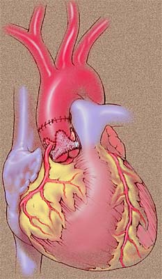
Implantation of a stentless xenograft aortic valve is technically more difficult than a stented valve but easier than an allograft used in the subcoronary position. Two valves approved for use by the United States Food and Drug Administration are the Toronto SPV (St. Jude, Minneapolis, MN) and the Freestyle valve (Medtronic, Minneapolis, MN). The Toronto SPV is comprised of the valve and supporting aortic wall only, and is designed as a subcoronary implant. The Freestyle valve is a root xenograft that may be used for either a subcoronary, cylindrical or a root replacement implant.

Operative Steps

A technique for the subcoronary implantation technique of the Toronto SPV is described.

Incision

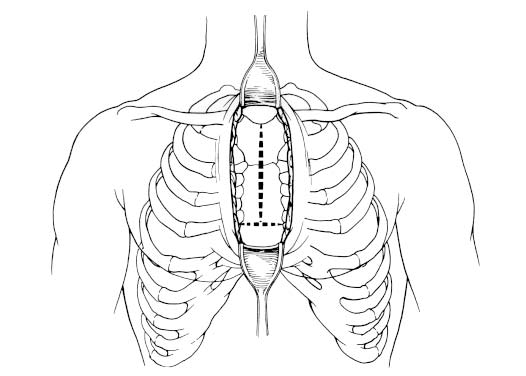
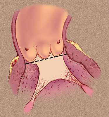
A full median sternotomy or upper hemisternotomy may be used. The upper hemisternotomy extends from the sternal notch to the level of the fourth intercostal space. At the inferior aspect of the upper hemisternotomy, the sternum can be partially or fully transected (Figure 1).

This incision permits adequate visualization of the cardiac structures to allow central cannulation with standard equipment (Figure 2).

Cardiopulmonary Bypass

Transesophageal echocardiography (TEE) is employed. A single two-stage venous cannula is placed in the right atrial appendage. The arterial cannula is placed in the distal ascending aorta. Both cannulas are connected to the cardiopulmonary bypass circuit. A retrograde cardioplegia cannula is inserted into the coronary sinus through a stab incision in the right atrium. The antegrade cardioplegia cannula is placed in the proximal ascending aorta. The right superior pulmonary vein is used for placement of the left ventricular vent. Dissection of the ascending aorta away from the pulmonary artery is performed. Cardiopulmonary bypass is established, a crossclamp is applied to the ascending aorta for administration of antegrade and retrograde cold blood cardioplegia.

Replacement Technique

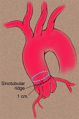
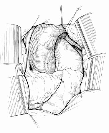
The aorta is opened transversely. It is essential to make this incision at least 1 cm above the sinotubular ridge. If the aorta is opened too close to the sinotubular ridge the valve posts will extend beyond the aortotomy, making proper commissural suspension difficult. The ascending aorta is completely transected (Figure 3).

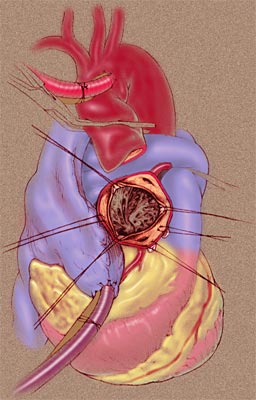
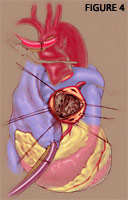
Exposure is obtained by elevating the valve annulus with sutures at each commissure and pledgeted 4-0 polypropylene sutures in the right and non-coronary cusps (Figure 4).

The native aortic valve is excised. The aortic annulus and aortic wall of the distal suture line are thoroughly debrided. The diameters of the annulus and sinotubular junction are measured. The size of the replacement valve is the larger of these two measured diameters as long as the difference in the measured diameters is less than 10%. Undersizing will result in valvular incompetence. If the difference in measured diameters is > 10%, consider using a porcine stentless or homograft as a root replacement, or a stented valve.

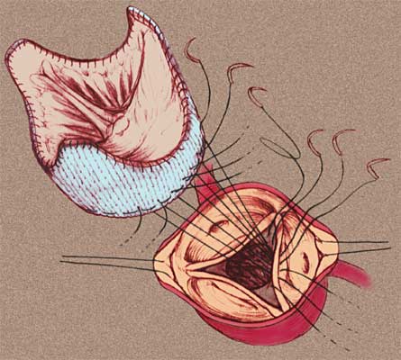
The muscular portion of the stentless xenograft valve (porcine right cusp) should be aligned with the non-coronary cusp region. The proximal (lower) suture line is placed in the sub-annular position with interrupted 4-0 Ticron sutures or a running 4-0 polypropylene suture. We use the interrupted technique (Figure 5).

The lower suture line is placed in the sub-annular position as a single horizontal plane (Figure 6).

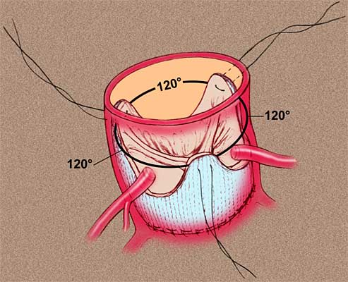
After tying the proximal suture line, each commissural post of the bioprosthesis is held in place with a single horizontal mattress 4-0 polypropylene suture. We do not tie these stay sutures until later. This maneuver ensures proper positioning of the commissural posts to achieve excellent coaptation of the aortic cusps and avoids occlusion of the coronary ostia. (Figure 7).

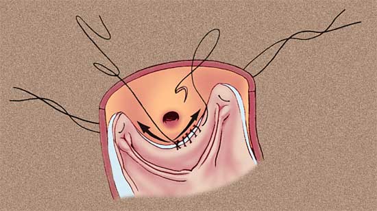
The distal suture line is a running 4-0 polypropylene suture that extends from the nadir of each cusp outward to the tip of each commissural post. It is this distal suture line that bears the stress of the valve closure. Care is taken to avoid obstruction of the coronary ostia. A full thickness of aortic wall is sutured to the bioprosthetic valve to prevent paravalvular leak. The aortic wall is lifted and kept on tension and the valve is pushed down while placing this suture line. This positions the valve to rest smoothly and firmly against the aortic wall, preventing gaps once the aorta is distended with blood (Figure 8).

The aortotomy is closed with two rows of a running 4-0 polypropylene suture (Figure 9).

De-airing is performed followed by removal of the aortic crossclamp. The patient is gradually wean off cardiopulmonary bypass, then decannulation is effected. Mean aortic transvavular gradient and coaptation of aortic cusps are documented with TEE. Atrial and ventricular wires are placed. After hemostasis is achieved, the sternum, fascia and skin are closed.

Preference Card

  • Set up for standard aortic valve replacement including left ventricular venting.
  • Have coronary ostial cannula available for antegrade cardioplegia.
  • Sutures include 3-0 or 4-0 Ticron with a RB-1 needle for the proximal suture line, 4-0 polypropylene with a RB-1 needle for the distal suture line, and 4-0 polypropylene with a SH-1 needle for the aortotomy closure.

Tips & Pitfalls

  • Avoid opening the aorta too proximal to the sinotubular junction. This leads to technical difficulty in closing the aorta, as the valve posts will extend beyond the aortotomy.
  • As with any aortic valve replacement, complete debridement of the diseased aortic valve, annulus and distal suture line is imperative.
  • Selection of stentless valve size is based on the sinotubular junction and annular diameters. The larger of the two diameters is the appropriate size as long as the diameters are within 10% of each other.
  • The muscular portion of the stentless xenograft valve should be aligned with the non-coronary cusp region.
  • The proximal suture line is placed in the sub-annular position as a single horizontal plane.
  • Suture bites in the muscle below the commissure between the right and noncoronary cusp should be shallow to avoid the Bundle of His.
  • Be sure valve posts are 120° apart, the same height and not twisted.
  • Keep 2 to 4 mm away from the coronary ostia.

References

  1. Reardon MJ, David TE. Stentless xenograft aortic valves. Curr Opin Cardiol 14: 84-89, 1999.
  2. David TE, Puschmann R, Ivanov J, Bos J, Armstrong S, Feindel CM, Scully HE. Aortic valve replacement with stentless and stented porcine valves: a case-match study. J Thorac Cardiovasc Surg 116:236-41, 1998.
  3. David TE, Feindel CM, Scully HE, Bos J, Rakowski. Aortic valve replacement with stentless porcine aortic valves: a 10-year experience. J Heart Valve Dis 7(3): 250-254, 1998.
  4. Reardon MJ, Conklin LD, Philo R, Letsou GV, Safi HJ, Espada R. The anatomical aspects of minimally invasive cardiac valve operations. Ann Thorac Surg 67: 266-68, 1999.

Add Comments Cancel reply

You must be logged in to post a comment.

Not a member yet? Register now.
TAGS
  • Valve Disease

Related Content

Article

Register for the Inaugural ‘In Africa for Africa’ Cardio-Thoracic Conference in Cape Town!

By: Marianna Mast

Video

ISMICS 2026 | Cardiac Session—Intraoperative Pearls and Pitfalls

By: Mario Castillo-SangBronze ContributorBronze Commentor

Podcast

The Beat With Joel Dunning Ep. 168: Humanoid Robots and AI in Surgery

By: Joel DunningGold ContributorSilver Commentor

Video

Robotic-Assisted Biventricular Durable Assist Device Implantation: The First In Human Experience

By: Feras Khaliel

Video

THVGreece 2026 | Tricuspid Valve

By: Evangelos Leontiadis

JANS Article

Humanoid Robots in the Operating Room: A Framework for Staged Integration of Embodied AI in Surgery

Author(s): Aamir Amin

JANS Article

Total Endovascular Repair of Ascending Aortic Pathology: The ENDOASC Multicentre Global Study

Author(s): Vincent SierSilver Contributor

JANS Article

Quality of Life After Cardiac Surgery: The QUACS Study and Benefit Prediction Model

Author(s): EACTS StaffSilver Contributor

Video

Management of SVC Injury During Right VATS Thymectomy for Myasthenia Gravis

By: Jakraphan Yu

Video

Single-Stage Repair of Left Ventricular Diverticulum with Ventricular Septal Defect Closure: A Video Illustration

By: Vatsal Vora

233 S. Wacker Drive | Suite 4400
Chicago, Illinois 60606, USA

Linkedin X-twitter Facebook-f Youtube
  • Join CTSNet
  • Donate
  • Submit Content
  • Volunteer
  • Join CTSNet
  • Donate
  • Submit Content
  • Volunteer
  • Featured Partners
  • Careers
  • Press
  • Advertise on CTSNet
  • Featured Partners
  • Careers
  • Press
  • Advertise on CTSNet
  • CTSNet Policies
  • Privacy Policy
  • Disclaimer
  • Contact CTSNet
  • CTSNet Policies
  • Privacy Policy
  • Disclaimer
  • Contact CTSNet

233 S. Wacker Drive | Suite 4400
Chicago, Illinois 60606, USA

Instagram Linkedin X-twitter Facebook-f Youtube
  • Join CTSNet
  • Donate
  • Submit Content
  • Volunteer
  • Join CTSNet
  • Donate
  • Submit Content
  • Volunteer
  • Featured Partners
  • Careers
  • Press
  • Advertise on CTSNet
  • Featured Partners
  • Careers
  • Press
  • Advertise on CTSNet
  • CTSNet Policies
  • Privacy Policy
  • Disclaimer
  • Contact CTSNet
  • CTSNet Policies
  • Privacy Policy
  • Disclaimer
  • Contact CTSNet

233 S. Wacker Drive | Suite 4400
Chicago, Illinois 60606, USA

Instagram Linkedin X-twitter Facebook-f Youtube
  • Join CTSNet
  • Donate
  • Submit Content
  • Volunteer
  • CTSNet Policies
  • Privacy Policy
  • Join CTSNet
  • Donate
  • Submit Content
  • Volunteer
  • CTSNet Policies
  • Privacy Policy
  • Featured Partners
  • Careers
  • Press
  • Advertise on CTSNet
  • Disclaimer
  • Contact CTSNet
  • Featured Partners
  • Careers
  • Press
  • Advertise on CTSNet
  • Disclaimer
  • Contact CTSNet
  • Join CTSNet
  • Donate
  • Submit Content
  • Volunteer
  • Join CTSNet
  • Donate
  • Submit Content
  • Volunteer
  • Featured Partners
  • Careers
  • Press
  • Advertise on CTSNet
  • Featured Partners
  • Careers
  • Press
  • Advertise on CTSNet
  • CTSNet Policies
  • Privacy Policy
  • Disclaimer
  • Contact CTSNet
  • CTSNet Policies
  • Privacy Policy
  • Disclaimer
  • Contact CTSNet
© 2026 CTSNet Inc. All right reserved.
Go to Top