Traumatic tracheobronchial injury is a rare but life-threatening complication of blunt chest trauma, accounting for approximately 0.5-2 percent of cases (1). These injuries most commonly occur near the right main bronchus due to the fixed position of the carina and reduced mediastinal protection (2). Prompt diagnosis and surgical intervention are essential to prevent respiratory failure and long-term complications.
This video demonstrates the case of a 44-year-old male who sustained a blunt chest injury after a heavy metallic plate fell directly onto his upper trunk. Upon initial evaluation, the patient exhibited tachycardia and hypoxemia, with an oxygen saturation of 88 percent on room air. Chest radiography revealed mediastinal emphysema, and an intercostal chest drain was placed. Subsequent contrast-enhanced computed tomography revealed a suspected right main bronchus injury associated with pneumomediastinum, pneumothorax, and fractures of the right second to fourth ribs.
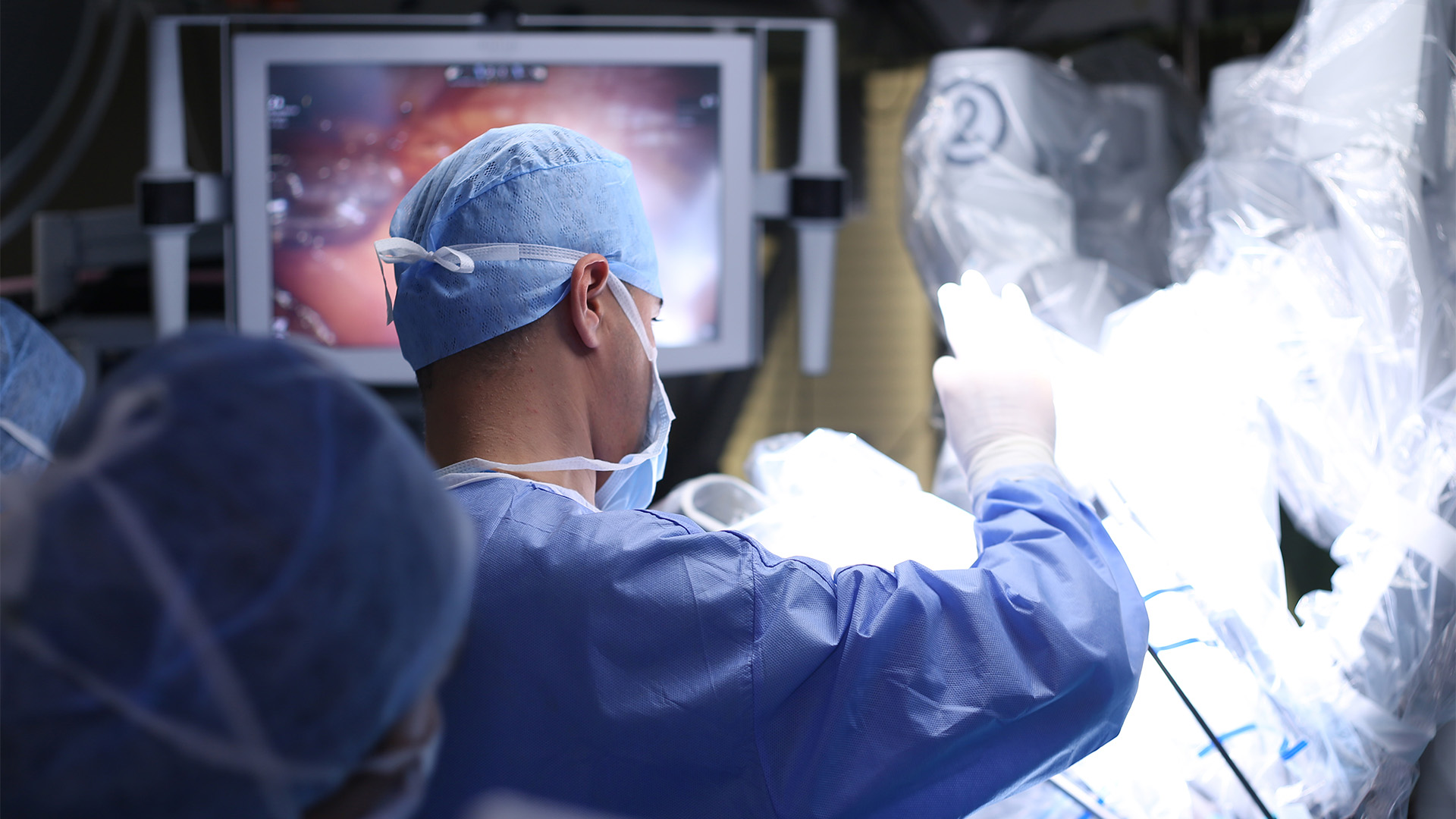
The patient was referred for emergency surgical management. Under general anesthesia with double-lumen endotracheal intubation, uniportal video-assisted thoracoscopic surgery (VATS) was performed through the right fourth intercostal space. Intraoperatively, a complete transection of the right main bronchus near the carina was identified. The azygos vein was divided to facilitate exposure of the injury site. Tracheal release maneuvers, including division of the inferior pulmonary ligament and mobilization of the right hilum, were performed to reduce anastomotic tension.
Reconstruction was achieved using polypropylene 4-0 sutures. The posterior membranous wall was repaired with a running suture, while the anterior wall was completed using interrupted sutures. The anastomosis was tested with underwater positive-pressure ventilation and reinforced with fibrin sealant and a pedicled pericardial fat pad buttress. Two chest tubes were placed for postoperative drainage.
The patient was extubated intraoperatively and transferred to the intensive care unit. Bronchoscopic evaluation on postoperative day three confirmed a patent airway and intact anastomosis. The chest tubes were removed on postoperative day three, and the patient was discharged on postoperative day seven without complications. Follow-up imaging demonstrated full lung expansion and no evidence of residual pneumothorax or airway stenosis.
Although thoracotomy remains the conventional surgical approach for traumatic bronchial injuries, this case demonstrates that uniportal VATS repair is feasible in selected patients. The minimally invasive approach offers potential advantages, including reduced postoperative pain, less tissue trauma, and improved visualization. Therefore, VATS may represent a safe and effective alternative to open thoracotomy in carefully selected cases of traumatic bronchial transection.
References
- Karmy-Jones R, Wood DE. Traumatic injury to the trachea and bronchus. Thorac Surg Clin. 2007;17:35-46.
- Cardillo G, Carbone L, Carleo F, et al. Tracheal lacerations after endotracheal intubation: a proposed morphological classification to guide non-surgical treatment. Eur J Cardiothorac Surg. 2010;37:581-587.
Disclaimer
The information and views presented on CTSNet.org represent the views of the authors and contributors of the material and not of CTSNet. Please review our full disclaimer page here.
 563 views on Vimeo