M1
 |
 |
|
Fig.
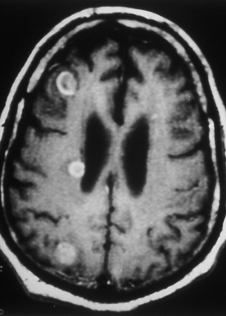
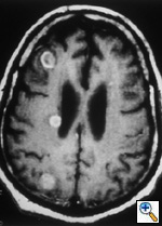
29: M1 disease.
Three enhancing metastases
of lung cancer are present
in this contrast enhanced
T1 magnetic resonance
image of the brain. |
|
|
M1 designates metastasis to distant organ
and lymph node sites. It is used also to
designate discontinuous tumor lesions outside
the parietal pleura in the chest wall or
in the diaphragm, and to classify metastasis
in ipsilateral non-primary tumor lobe(s).
 |
|
|
Fig.
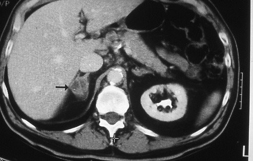
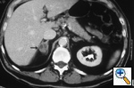
30: M1 disease.
A computed tomographic
scan of the abdomen shows
metastatic lung cancer
to the right adrenal
gland. The gland is enlarged
(arrow) and shows areas
of decreased attenuation
in keeping with necrosis. |
|
|
|