Stage
IIIB
The role of magnetic resonance imaging
for the evaluation of lung tumors has not
been fully developed; however, the technique
has been shown to be of particular value
for evaluating tumors arising in the superior
sulcus of the lung.
STAGE IV
The stage IV category includes
only patients with evidence of distant
metastasis,
M1 disease. Clinical signs and symptoms of metastasis to distant organ sites
usually are confirmed by imaging techniques; however, routine brain and bone
scans have not proved cost-effective for patients with non-small cell lung
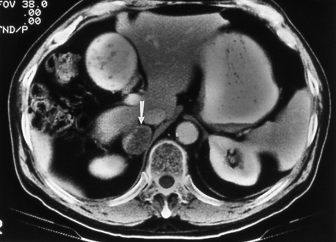
cancer. The significance of adrenal
nodules imaged on abdominal CT should
be determined
by biopsy. Other definitive signs and symptoms of distant metastasis need
not be biopsy proved for assigning the
M1 category. Positron emission tomography
(PET) holds promise for making clinical estimates of metastatic disease more
reliable; however, specific indications for its use and application of the
findings remain understudy13. Ipsilateral
metastasis in non-primary-tumor lobes
also
is designated M1-Stage IV (see pages 6 and 26).
 |
 |
|
Fig.
50: Computed
tomographic scan of the
abdomen showing a necrotic
metastasis in the right
adrenal gland (white
arrow) from a primary
bronchogenic carcinoma,
M1, stage IV. |
|
|
|