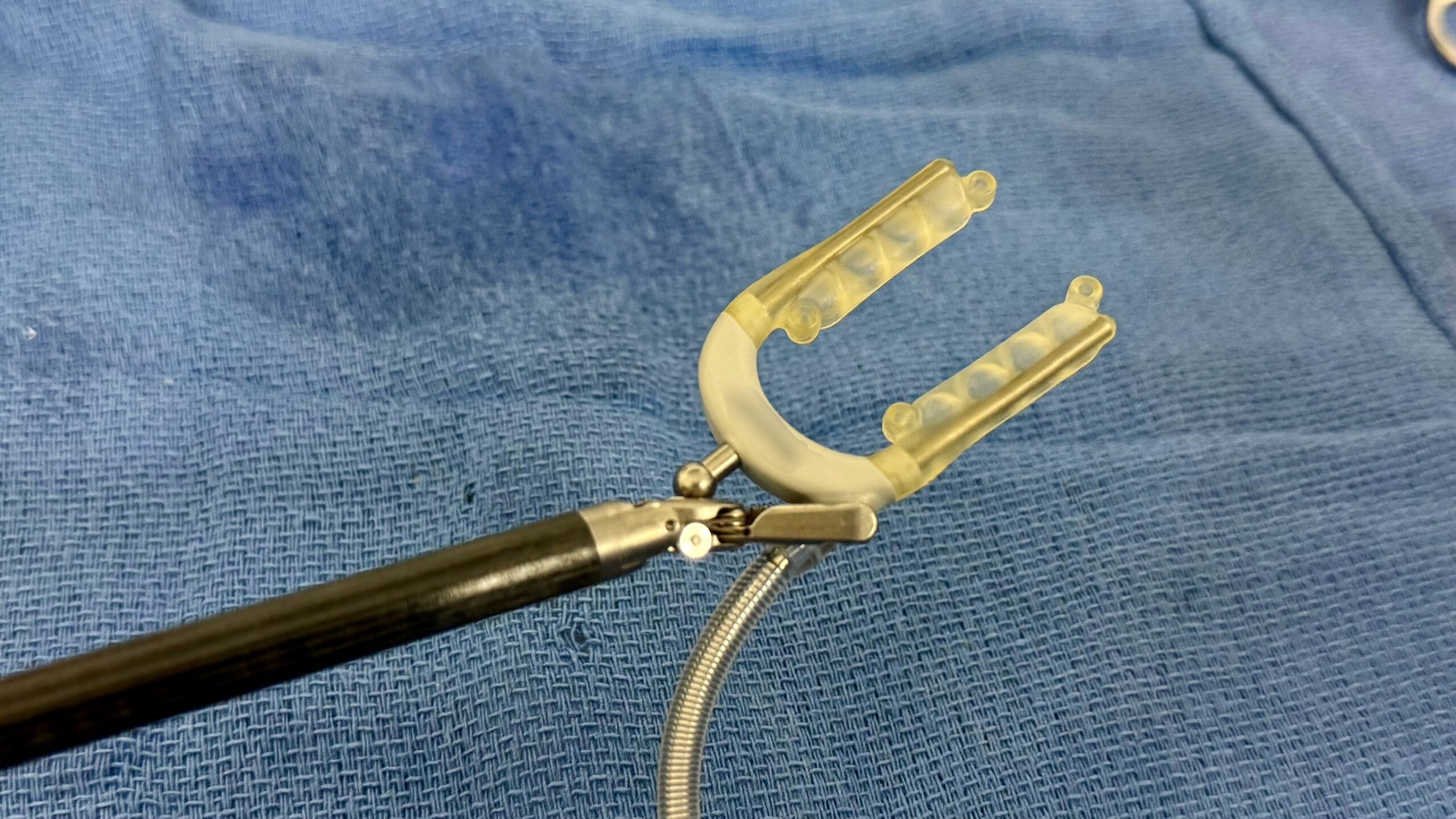
This video demonstrates skeletonized harvesting of the left internal thoracic artery (LITA) through a left anterior minithoracotomy.
Exposure and Initial Dissection
After the thoracotomy, careful exposure began with identification of the mammary course and proper placement of the retractor blade to avoid injury to the vessel during rib spreading. Although complete left lung deflation provides the best visualization, the procedure can also be performed safely with reduced lung inflation.
Dissection began at the first visible segment of the artery, where the overlying muscle and fascia were divided to expose the LITA within in a skeletonized plane. The harvest proceeded proximally until the subclavian vein was identified, with meticulous division of the mediastinal and first intercostal branches.
Graft Mobilization
In this technique, clips are deliberately avoided whenever possible to preserve a completely skeletonized graft while minimizing foreign material in the operative field. Particular attention is paid to separating the artery from its concomitant veins, especially the larger medial vein. This improves graft length and facilitates identification of proximal branches while reducing the risk of troublesome bleeding.
Following complete mobilization, the distal LITA was divided, papaverine was applied, and the operation proceeded according to the planned strategy.
Disclaimer
The information and views presented on CTSNet.org represent the views of the authors and contributors of the material and not of CTSNet. Please review our full disclaimer page here.
 2 views on Vimeo