The frozen elephant trunk (FET) technique provides a useful surgical approach for managing complex aortic arch pathologies (1). One of the critical challenges of this procedure is the reimplantation of the left subclavian artery (LSA) (2).
A new hybrid device, the E-vita Neo EDE, (3) combines a standard FET prosthesis with an additional branch specifically designed for the open stenting of the LSA. Contraindications for this device may include a left vertebral artery origin distance of less than 4 mm, a separate origin of the vertebral artery, aneurysms, stenosis, or significant kinking of the LSA (4).
A 67-year-old male patient who had been under surveillance for two years underwent zone 0 FET implantation with this novel, custom-designed hybrid device.
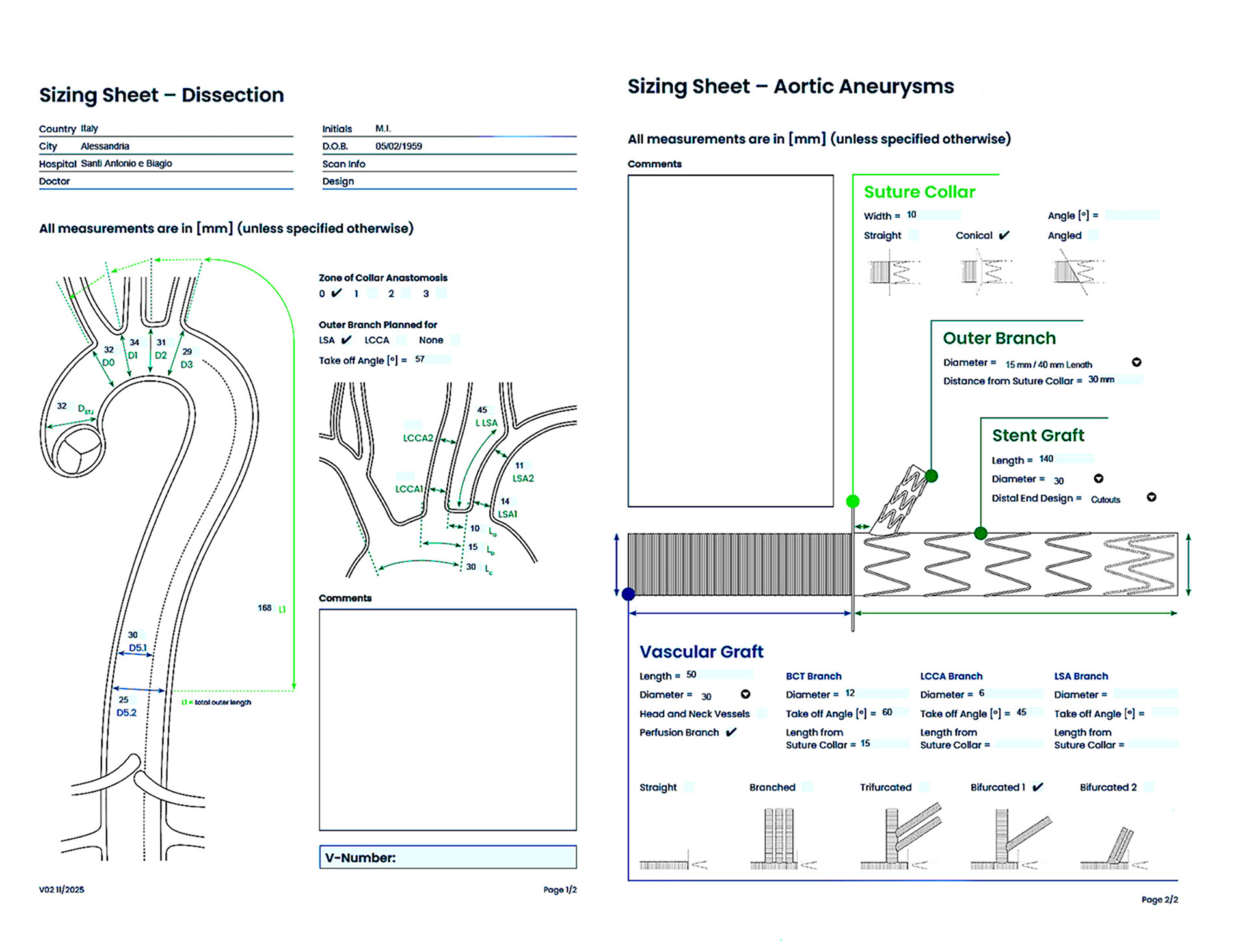
Figure 1: The sizing sheet for aortic aneurysms and dissection repair.
The computed tomography (CT) scan showed a residual dissection, a partially thrombosed false channel with arch and descending aortic dilatation. The 3D reconstruction highlighted compression of the true lumen, false channel extension, and involvement of the visceral vessels.
Before sternotomy, a guidewire was advanced into the left subclavian artery through the brachial artery. Simultaneous transesophageal echocardiography showed preserved biventricular function and trivial aortic valve regurgitation.
The median sternotomy was extended to the right neck. The innominate artery was exposed, dissected up to the bifurcation, and encircled with an umbilical tape. In the same fashion, the left carotid artery was prepared. Two simple box-shaped stitches 5-0 polypropylene placed side-by-side in opposite directions and buttressed with the Teflon-felt pledgets were placed on the anterior wall of the proximal portion of the right subclavian artery. The subclavian artery was directly cannulated for cardiopulmonary bypass (CPB).
After the right atrial appendage cannulation and the left ventricle venting through the right superior pulmonary vein, CPB and systemic cooling to approximately 28°C were started. The right carotid artery was separated using a surgical stapler, and an 8 mm Dacron graft was sutured in an end-to-end fashion to the artery and cannulated via a Y-line from the arterial line.
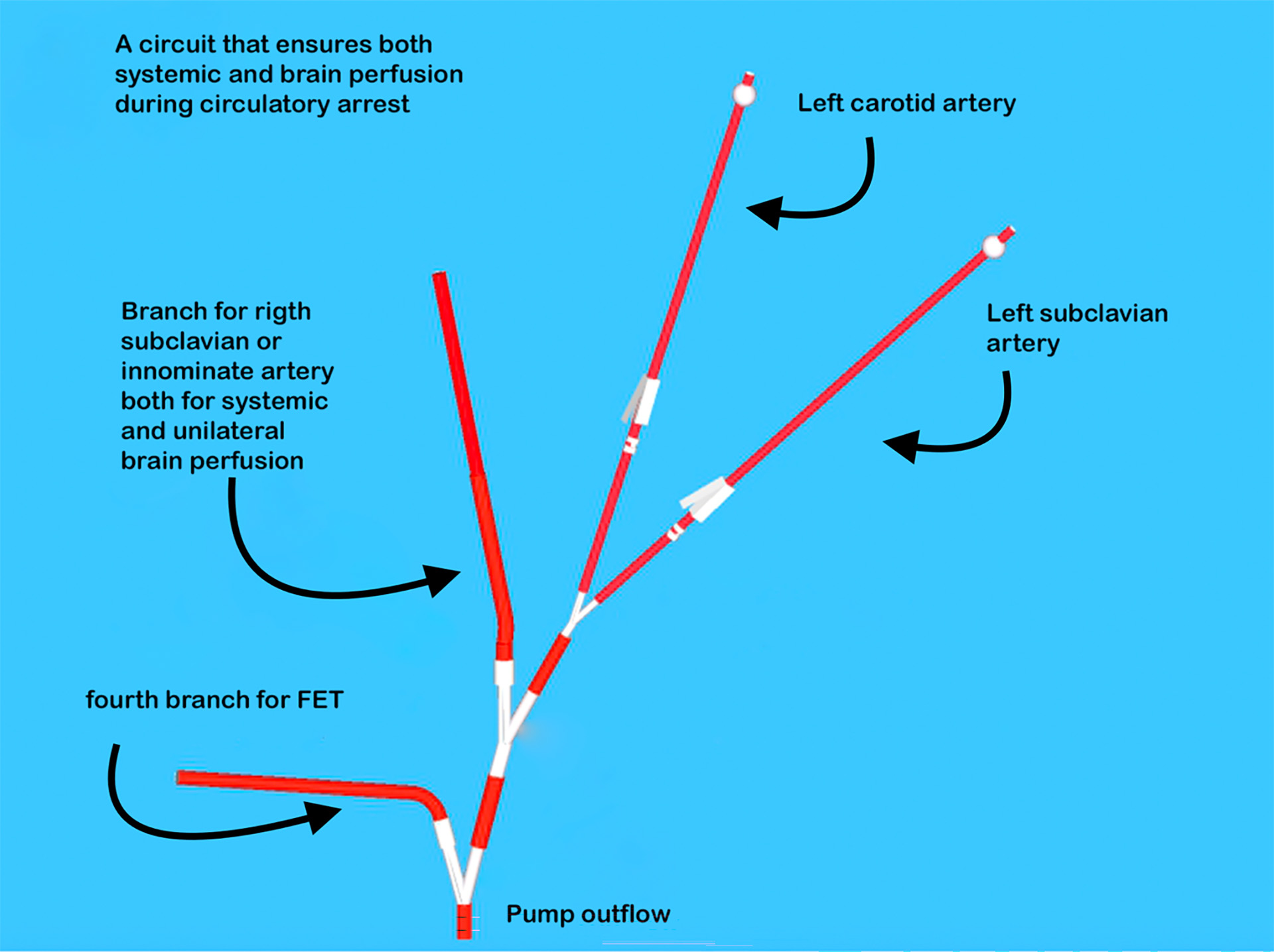
Figure 2: Circuit for brain and systemic perfusion.
Cerebral perfusion was monitored by near-infrared spectroscopy (NIRS). Spinal drainage is not routinely performed preoperatively unless a distal FET landing below the eighth thoracic vertebra (T8) is expected. The graft previously implanted was carefully isolated and cross-clamped just proximal to the innominate artery.
Myocardial preservation was achieved through the infusion of cold histidine-tryptophan-ketoglutarate (HTK) solution, administered in an antegrade fashion. At 28°C, the CPB flow was stopped, the innominate artery was clamped, the aortic cross-clamp was removed, and selective bilateral antegrade brain perfusion was started at 10 mL/kg/min.
A transverse incision was performed across the graft. The next step involved the introduction of guidewires into the main body of the prosthesis and subsequently into the subclavian stent branch. The E-vita Neo EDE was gently pushed into the descending aorta via the guidewire; the FET stent was released, followed by the side branch stent in the LSA and the guidewires were removed.
During the distal anastomosis between the aorta and the FET collar, a Foley catheter was placed into prosthesis, and reperfusion to the lower body was started.
By performing the proximal anastomosis soon after the distal anastomosis, the cardiac ischemic time can be reduced. The Foley catheter was positioned in the fourth branch.
After careful deairing, systemic perfusion and rewarming were initiated through the fourth branch. Perfusion of the left common carotid was then stopped, and the artery was anastomosed to the corresponding side branch.
Finally, the innominate artery was clamped and, while being perfused, was anastomosed to its corresponding side branch. The sutures were reinforced with autologous pericardium.
Systemic rewarming, decannulation, meticulous hemostasis, and closure of the thorax and wound were completed without complications. The patient was transferred to the intensive care unit in stable hemodynamic condition. The patient regained consciousness seven hours after surgery and was weaned from mechanical ventilation on postoperative day one. The postoperative course remained uneventful, and the patient was discharged on postoperative day eight.
The predischarge CT angiography showed no residual flow in the false lumen across the arch and descending aorta, with normal flow in the innominate, right subclavian, right carotid, and left carotid arteries, as well as regular perfusion of the LSA.
Conclusion
The hybrid arch device streamlined FET implantation, offering a user-friendly solution that facilitated surgical management of the LSA without increasing procedural complexity.
References
- Weiss G, Arnold Z, Folkmann S, Aschacher T, Tauber S, Harrer M L et al. From back table innovation to contemporary application: a review of the frozen elephant trunk technique.Ann Cardiothorac Surg 2025;14(5):335-342
- Orozco-Sevilla V, Coselli JS. Management of the left subclavian artery during aortic arch replacement using a frozen elephant trunk approach: a review. Cardiovasc Diagn Ther 2023;13:736–42.
- Folkmann S, Arnold Z, Geisler D, Lenz V, Miosga D, Harrer M et al. First-in-men experience with a novel frozen elephant trunk prosthesis featuring an endovascular side branch for left subclavian artery connection. Eur J Cardiothorac Surg 2024;66:ezae302.
- Pitts L, Kofler M, Montagner;M, Kempfert J. Total Arch Replacement with a New Hybrid Device to Manage the Left Subclavian Artery in the Frozen Elephant Trunk Technique. Interdisciplinary CardioVascular and Thoracic Surgery 2026, 41(3)
Disclaimer
The information and views presented on CTSNet.org represent the views of the authors and contributors of the material and not of CTSNet. Please review our full disclaimer page here.
 549 views on Vimeo