This video is part of CTSNet’s 2025 Resident Video Competition. Watch all entries into the competition, including the winning videos.
Diaphragm Pacing (DP)
DP electrically stimulates the diaphragm at phrenic nerve motor end points to stimulate diaphragm contraction, improving and preserving diaphragm function in patients with phrenic nerve injury (PNI) and diaphragm dysfunction (DD).
Indications
Early investigations reserved DP for patients who were ventilator-dependent due to high spinal cord injury and central hypoventilation syndrome. However, recent advances in DP have expanded its use to select patients with phrenic nerve injury and diaphragm dysfunction, including those who have undergone cardiac surgery or heart and lung transplants.
Pacing Goals
For patients with cervical spinal cord injury who are dependent on mechanical ventilation, the primary goal is to wean them from the ventilator, achieving a minimum of four continuous hours of ventilator-free time. This intervention also aims to avoid ventilator-induced lung injury (VILI).
In patients with phrenic nerve injury and diaphragm dysfunction, the goal of DP include accelerating recovery of PNI, preserving diaphragmatic function, preventing diaphragm atrophy, and alleviating symptoms.
Stimulation Physiology
In DP, an external pulse generator provides repeated twitches that trigger repeated action potentials. The frequency of these pulses “fuses” the contractions, creating a state of tetany. The principle is used in DP mapping, where more propagated action potentials are recruited closer to the phrenic nerve trunk.
Respiration
The simulation involves repeated trains of pulses that correspond to the inspiratory contraction phase. This results in a cycle of repeated diaphragmatic contractions and relaxations. The respiratory rate is determined by the frequency of train repetition.
Preoperative Considerations
The phrenic nerve must be intact, whether fully or partial, for successful DP. This can be evaluated using fluoroscopy with diaphragm stimulation (the sniff test), diaphragm ultrasonography, and diaphragm conduction studies. Additionally, the diaphragm must respond to electrical stimulation. Direct stimulation (mapping) is usually done at the same time as DP, but it can also be performed during a prior diagnostic laparoscopy.
Preoperative Workup
A complete cardiopulmonary workup is necessary to exclude other causes of symptoms. This workup includes an echocardiogram, stress test, and cardiac catheterization. Pulmonary function tests, perfusion scans, and cardiopulmonary exercise testing are also part of the evaluation. Cross-sectional imaging may be performed as well.
Diaphragm Pacer and Mapping
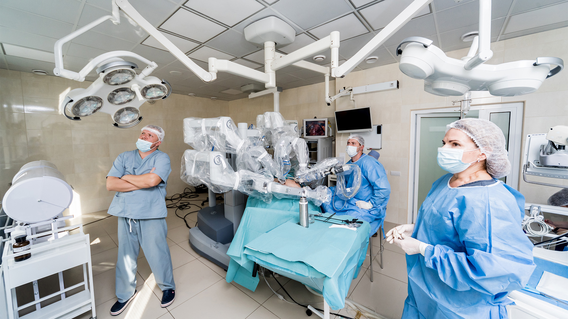
DP involves robotically placing four electrodes, with two in each hemi-diaphragm, at the phrenic nerve motor end plate areas that control diaphragm contractions. The electrodes are tunneled under the skin to an exit site. These electrodes connect to the battery-powered external pulse generator (EPG), which is programmed to stimulate the muscle and the phrenic nerves to cause diaphragm contraction. Direct motor end point stimulation is conducted to identify the area with maximum contraction.
Brief Case Summary
A 71-year-old male presented with a 10-month history of severe exertional dyspnea requiring supplemental oxygen. He had a history of COVID infection, hypertension, hyperlipidemia, obstructive sleep apnea, gastroesophageal reflux disease (GERD), and a remote coronary artery bypass grafting (CABG).
Workup revealed an elevated, paralyzed right hemidiaphragm on chest X-ray (CXR) and fluoroscopy. An extensive cardiopulmonary workup revealed no other source of dyspnea. The patient was counseled for right diaphragm stimulator placement, and an u
neventful robotic diaphragm pacer mapping and placement was performed. The pacer units were programmed for comfort, and he was discharged home on postoperative day one.
At the three- and six-months follow-up, the patient reported significant symptom relief and no longer required oxygen. The device was reprogrammed to higher amplitudes. The patient provided the following testimonial:
“I can now go about my regular daily activities without being short of breath and needing oxygen.” Future follow-up will assess phrenic nerve recovery.
References
- Giberson CE, Cheshier SH, Poree LR, Saulino MF. Diaphragm Pacing: A Safety, Appropriateness, Financial Neutrality, and Efficacy Analysis of Treating Chronic Respiratory Insufficiency. Neuromodulation. 2023 Apr;26(3):490-497. doi: 10.1016/j.neurom.2022.10.059. Epub 2023 Jan 4. PMID: 36609087.
- Cavka K, Fuller DD, Tonuzi G, Fox EJ. Diaphragm Pacing and a Model for Respiratory Rehabilitation After Spinal Cord Injury. J Neurol Phys Ther. 2021 Jul 1;45(3):235-242. doi: 10.1097/NPT.0000000000000360. PMID: 34049339.
- Kerwin AJ, Zuniga YD, Yorkgitis BK, Mull J, Hsu AT, Madbak FG, Ebler DJ, Skarupa DJ, Shiber JR, Crandall ML. Diaphragm pacing improves respiratory mechanics in acute cervical spinal cord injury. J Trauma Acute Care Surg. 2020 Sep;89(3):423-428. doi: 10.1097/TA.0000000000002809. PMID: 32467474.
- Kerwin AJ, Yorkgitis BK, Ebler DJ, Madbak FG, Hsu AT, Crandall ML. Use of diaphragm pacing in the management of acute cervical spinal cord injury. J Trauma Acute Care Surg. 2018 Nov;85(5):928-931. doi: 10.1097/TA.0000000000002023. PMID: 29985232.
- Onders RP, Elmo M, Kaplan C, Schilz R, Katirji B and Tinkoff G. Long-term experience with diaphragm pacing for traumatic spinal cord injury: Early implantation should be considered. Surgery. 2018;164:705-711.
- Posluszny JA, Jr., Onders R, Kerwin AJ, Weinstein MS, Stein DM, Knight J, Lottenberg L, Cheatham ML, Khansarinia S, Dayal S, Byers PM and Diebel L. Multicenter review of diaphragm pacing in spinal cord injury: successful not only in weaning from ventilators but also in bridging to independent respiration. J Trauma Acute Care Surg. 2014;76:303-9; discussion 309-10.
Disclaimer
The information and views presented on CTSNet.org represent the views of the authors and contributors of the material and not of CTSNet. Please review our full disclaimer page here.
 392 views on Vimeo
 1,347 views