


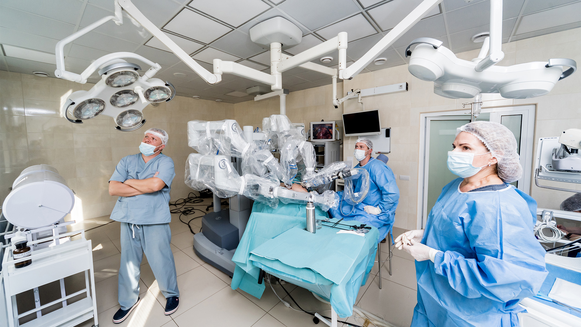
This video demonstrates the robotic resection of a giant superior mediastinal mass. The patient was a middle-aged male who had been referred to the authors’ clinic for evaluation of a mediastinal mass discovered on magnetic resonance imaging (MRI) during workup for dorsalgia. Imaging originally demonstrated a large, approximately 7 cm mass in the right upper mediastinum associated with tracheal deviation.
Further workup with computed tomography (CT) of the chest demonstrated similar findings and a small, central, coarse calcification. Positron emission tomography (PET)/CT demonstrated minimal avidity, most suggestive of a nerve sheath tumor. This patient was scheduled for a flexible bronchoscopy, endobronchial ultrasound, and transbronchial needle aspiration for a total of five biopsies, all of which were nondiagnostic. Cultures, however, were positive for Schaalia odontolytica, and the patient was started on Augmentin. He was referred to Infectious Disease, who recommended continuing antibiotics and obtained a three-month interval CT scan. If no change was appreciated, they recommended resection of the mass for diagnostic and therapeutic purposes. The three-month interval CT scan demonstrated a large mass, relatively unchanged in appearance. There was also persistent tracheal narrowing. Due to mass effect, especially on the trachea, the patient was offered right robotic-assisted thoracoscopic resection of the mediastinal mass.
The mass was located in the superior mediastinum, above the azygos vein, between the trachea, superior vena cava, and subclavian artery. The pleura overlying the mass was divided along the anterior border, taking care to spare the recurrent laryngeal nerve and any critical surrounding structures. A 2-0 Ethibond suture was attempted to be placed in a figure-of-eight fashion to help with retraction and dissection along the anterior border of the mass. However, significant progress with the dissection was difficult to achieve. A sample of the mass was taken using bipolar forceps and sent to pathology. Intraoperatively, the pathology returned as nondiagnostic. Additional 2-0 Ethibond sutures were placed at different points of the mass to aid in retraction.
Because the prior pathology had been nondiagnostic, the mass was transected at the posterior portion using monopolar scissors for further sampling. The mass itself appeared white and tan. The divided mass was sent for pathology, which returned as a mesenchymal tumor. Additional retraction sutures were placed facilitate anterior dissection. During the dissection, blunt dissection was primarily used, allowing maintenance of a plane directly on top of the mass capsule. This avascular plane allowed for safely dissection without damaging critical surrounding structures and vasculature. Finally, the mass was freed from all surrounding tissue and adhesions. The entire mass was removed from the superior mediastinum and was sent to pathology.
Postoperative chest x-ray showed no pneumothorax or pleural effusion and a lack of significant mediastinal pathology. Additionally, a chest tube was seen in the right chest, which was removed on postoperative day one without issue. Final pathology demonstrated myxolipomatous neoplasm, favoring pleomorphic lipoma.
Pleomorphic lipoma is a soft tissue mass usually found in the subcutaneous tissues of the neck or shoulders in older males. Grossly, it resembles a normal lipoma, but histologically, it is defined by pleomorphic multinucleated giant cells. Because of this, it is often confused with liposarcoma on pathology. However, pleomorphic lipoma is a benign tumor, where excision is usually curative.
This video submission is from the 2026 CTSNet Instructional Video Competition. Stay tuned to CTSNet.org and the CTSNet YouTube channel in the coming weeks to watch all entries from the competition, including the winning videos.
References
- Sakhadeo U, Mundhe R, DeSouza MA, Chinoy RF.Pleomorphic lipoma: A gentle giant of pathology. J Cytol.2015 Jul-Sep;32(3):201-3. doi: 10.4103/0970-9371.168904. PMID: 26729985; PMCID: PMC4687215.
- Shmookler BM, Enzinger FM. Pleomorphic lipoma: a benign tumor simulating liposarcoma. A clinicopathologic analysis of 48 cases. Cancer. 1981 Jan 1;47(1):126-33. doi:10.1002/1097-0142(19810101)47:1<126:aid-cncr2820470121>3.0.co;2-k. PMID: 7459800.
- Ohshima Y, Nishio J, Nakayama S, Koga K, Aoki M, YamamotoT. Spindle Cell Lipoma and Pleomorphic Lipoma: An Update and Review. Cancer Diagn Progn. 2023 May 3;3(3):282-290. doi: 10.21873/cdp.10213. PMID: 37168965; PMCID:PMC10165376.
Disclaimer
The information and views presented on CTSNet.org represent the views of the authors and contributors of the material and not of CTSNet. Please review our full disclaimer page here.
 238 views on Vimeo
 396 views












1 Comment
Very interesting case .
Thank you