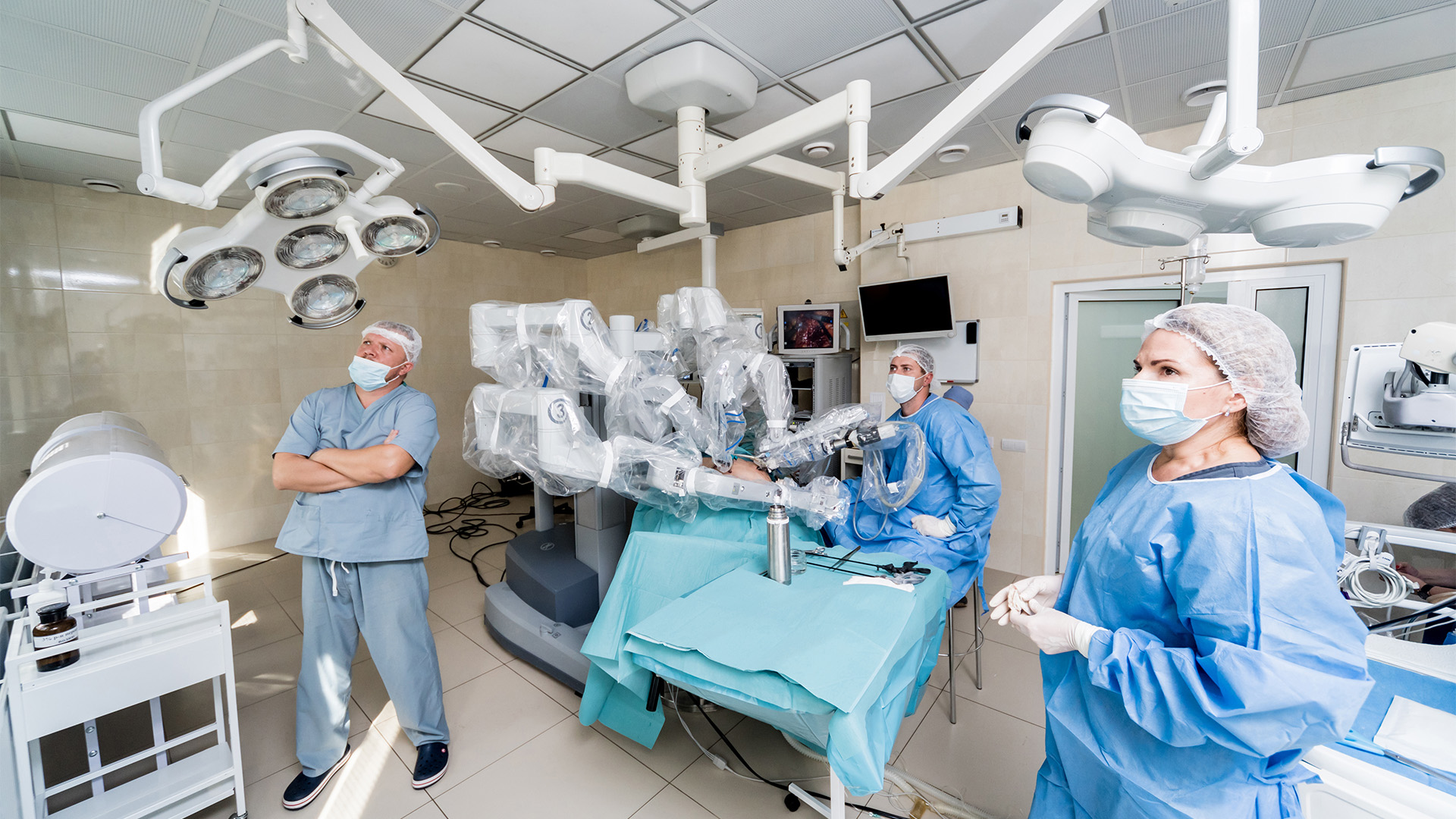
The video begins with an interview featuring CTSNet Senior Editor—Education Vincent Gaudiani, a Clinical Associate Professor of Cardiothoracic Surgery at Stanford University in Stanford, California, USA, and Lorena Montes, a cardiothoracic surgeon at Fundacion Cardiovascular de Colombia in Floridablanca, Colombia, interviewing the author of this complex case Oleksandr Babliak, Chief of Cardiac Surgery Department, Cardiac Surgery Center, Dobrobut Medical Network, Kyiv, Ukraine.
They explore the challenges of this case and explain the decision-making process behind the surgical techniques that were employed. After the interview, the surgical case is presented.
Case
The authors present a case demonstrating the surgical management of a patient with a porcelain aorta undergoing combined aortic valve replacement, mitral valve repair, and coronary artery bypass grafting.
A 68-year-old woman presented with Canadian Cardiovascular Society (CCS) class III–IV angina, dyspnea on exertion, presyncope, and dizziness. Transthoracic echocardiography revealed severe low-flow, low-gradient aortic stenosis (aortic valve area [AVA] 0.7 cm²), severe mitral regurgitation (Carpentier type IIIa), severe pulmonary hypertension, and a reduced left ventricular ejection fraction (LVEF) of 32 percent. Her past medical history included radiation therapy for Hodgkin lymphoma more than 30 years ago, right coronary artery (RCA) stenting in 2014, left anterior descending (LAD)-left circumflex (Lcx) bifurcation stenting in 2018, and atrial fibrillation ablation in 2019. Preoperative whole-body computed tomography (CT) angiography demonstrated a porcelain aorta and significant stenoses of the brachiocephalic and subclavian arteries. Coronary angiography confirmed significant disease of the obtuse marginal, left anterior descending, and posterior descending arteries. The previously implanted RCA ostial stent was protruding into the aortic root, which created additional concerns regarding future catheter-based procedures.
The patient required surgical coronary revascularization due to recurrent restenosis and progressive coronary disease despite prior interventions.
A porcelain aorta presented a major technical challenge due to the high risk of embolic complications associated with aortic manipulation; therefore, a carefully tailored operative strategy was adopted. The procedure was performed via median sternotomy with peripheral cannulation. Under reduced flow, a transthoracic aortic cross-clamp was applied, and an aortotomy was performed.
Due to the protrusion of a previously implanted RCA stent, antegrade cardioplegia delivery through the coronary ostia was not feasible, and myocardial protection was achieved using retrograde cardioplegia. Following the excision of the aortic valve, extensive decalcification of the ascending aorta was undertaken. To facilitate complete and safe decalcification, the patient was cooled to 28°C, and a short period of circulatory arrest was initiated. The circulatory arrest time was limited to eight minutes, which allowed for the effective removal of calcific plaques and restoration of a clampable aortic segment.
The left internal mammary artery was intentionally avoided because of the significant left subclavian artery stenosis. Coronary revascularization was performed using a saphenous vein graft for sequential bypass to the posterior descending artery, the obtuse marginal branch, and the left anterior descending artery. The mitral valve anatomy and the need for a durable result favored surgical repair over edge-to-edge transcatheter treatment. The mitral valve repair was completed using an annuloplasty band, and a 21 mm bioprosthetic valve was implanted in the aortic position.
The aortotomy was closed with 5-0 polypropylene sutures, and proximal vein graft anastomoses were performed during the cross-clamp period. After meticulous deairing, the aortic cross-clamp was removed.
The postoperative course was uneventful, and the patient was discharged home without neurological complications. Regarding long-term results with aortic endarterectomy, at the two-year follow-up, the patient remained free of neurological deficits, aortic enlargement, or acute aortic syndrome. Echocardiography performed two years postoperatively showed an ascending aortic diameter of 3.0 cm.
Disclaimer
The information and views presented on CTSNet.org represent the views of the authors and contributors of the material and not of CTSNet. Please review our full disclaimer page here.
 543 views on Vimeo
 931 views