Patient Selection
Ligation of the thoracic duct is indicated in any patient with a traumatic or iatrogenic chylothorax who has not responded to a brief trial of conservative management such as a low-fat diet, medium-chain triglycerides, total parenteral nutrition, and/or octreotide [1]. Spontaneous chylothorax, usually associated with mediastinal diseases such as lymphoma, are not often amenable to such therapy. Both open and thoracoscopic approaches to duct ligation are feasible in most patients. Chylothorax following minimally invasive pulmonary surgery is easily managed by VATS thoracic duct ligation. Chylothorax following minimally invasive esophagectomy either with a combined trans-thoracic trans-abdominal approach or a completely trans-abdominal approach is also an indication for thoracoscopic ligation of the duct [2]. In contrast, chylopericardium is managed by pericardial drainage and ligation of the duct, both of which can be performed thoracoscopically [3]. An unusual situation is presented by patients who have a thoracic duct cyst; they will require ligation of the duct during excision of the cyst [4].
In patients who have had a recent thoracotomy (within 7-10days) there is no advantage in performing thoracoscopy to ligate the duct. The thoracotomy should be reopened if ligation of the duct is required, particularly if the thoracotomy has been performed on the right. Thoracoscopic ligation of the thoracic duct may still be appropriate if the thoracotomy which resulted in the chylothorax was performed on the left side.
Operative Steps
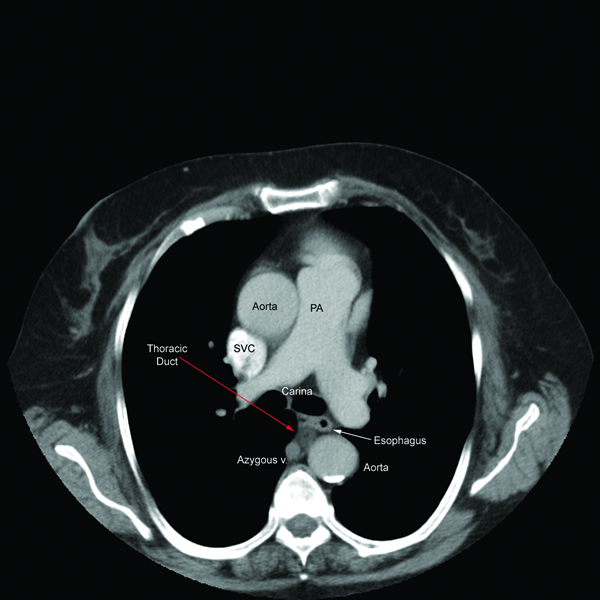
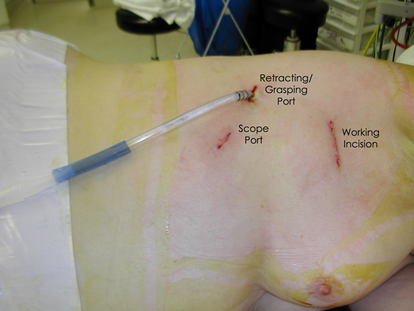
General anesthesia with single lung ventilation is required and the duct is always approached through the right chest due to its constant position in the lower right chest (Figure 1). The patient is placed in a left lateral decubitus position, rolled forward, and flexed to allow increased exposure of the posterior mediastinum. An epidural is not necessary and intercostal blocks with 0.25% bupivacaine can be used for analgesia. The thoracoscope is placed in the seventh intercostal space along the line of the anterior superior iliac spine and the working incision (5cm) is placed in the fifth or sixth intercostal space in the anterior axillary line (Figure 2). The soft tissues of the chest wall are kept open with a Weitlaner retractor and the intercostal incision is made longer than the skin incision. This allows for the introduction of two or three instruments through this incision and allows suction to be applied within the chest without causing re-expansion of the lung. A third incision is made at the level of the dome of the diaphragm after visualizing the desired space thoracoscopically. This incision is placed along the mid-axillary line.
 |
 |
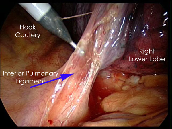 |
| Figure 1: CT Chest showing thoracic duct. | Figure 2: Thoracoscopic port placement. | Figure 3: Inferior Pulmonary Ligament Division. |
A 30 degree scope is used to visualize the chest and a tonsil sponge introduced through the incision over the diaphragm (Figure 2) is used to retract the diaphragm inferiorly. Simultaneously, the postero-inferior edge of the lower lobe is grasped with a ring clamp introduced through the working incision and retracted superiorly and posteriorly to display the inferior pulmonary ligament, which is then divided with cautery (Figure 3). A Yankauer sucker placed through the working incision allows for a clear field unobstructed by cautery smoke. Care must be taken as the inferior pulmonary vein is approached and it is preferable to use scissors to mobilize this part of the ligament. Gentle blunt dissection with a tonsil sponge or peanut is helpful in completing the dissection and allowing the inferior pulmonary vein to be identified. This dissection is then continued either sharply or with cautery, superiorly for a distance, opening the mediastinal pleura posterior to the hilum until the bronchus intermedius is reached. This allows the lung to be retracted anteriorly with a tonsil sponge or fan retractor which increases the exposure to the posterior mediastinum. Rolling the operating table forward further facilitates this exposure.
The posterior mediastinal pleura between the azygous vein and the chest wall is initially opened and the vein is mobilized. This allows the vein to be retracted without avulsion of intercostal venous tributaries (Video 1). The mediastinal pleura between the azygous vein and the esophagus is opened longitudinally with cautery or scissors (Video 2). This is done at a point inferior to the level of the inferior pulmonary vein, as at this level the duct is most often solitary and has not duplicated, which may occur at a higher level. The esophagus is retracted anteriorly using a ring clamp or sponge stick and the dissection continued bluntly towards the aorta using a peanut dissector (Videos 3, 4). Venous tributaries from the azygos vein and arteries from the aorta to the esophagus can be clipped with a 5mm clip applier and divided sharply. Alternatively, they can be divided with a harmonic scalpel.
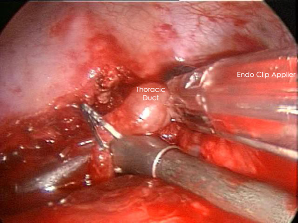
Exploration of this area bluntly will reveal the thoracic duct as a thin tubular structure with occasional peristalsis (Video 5). Once a sufficient length of duct is isolated it is doubly clipped using 5 or 10mm clips depending on the size of the duct and is divided (Figures 4-6). Transection with a linear cutting stapler with a vascular load (30mm length, 2.0mm staple height) works just as well (Video 6). The stapler or clip applier can be introduced through the working incision or the incision used for retraction of the diaphragm. If an adequate length of the duct can be isolated, a portion can be excised and sent for frozen section to confirm its identity.
 |
 |
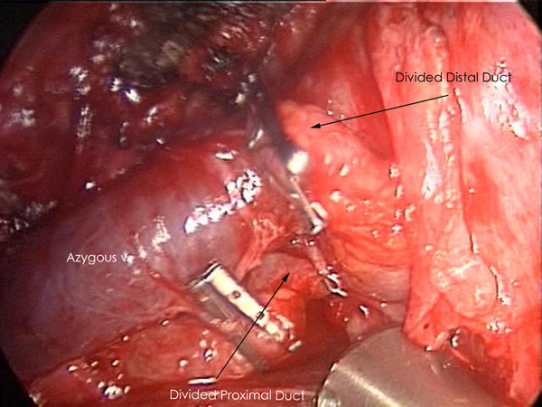 |
| Figure 4: Thoracic Duct isolation. | Figure 5: Thoracic Duct Ligation. | Figure 6: Thoracic Duct Divided. |
If the duct is not directly identified, all fatty and lymphatic tissue anterior to the azygous vein, posterior to esophagus, and between the aorta and right parietal pleura is divided between endoclips, intracorporeally placed ligatures, or with a linear cutting stapler with a vascular load. Since these are the anatomic bounds of where the thoracic duct is located, these maneuvers can successfully ligate the duct without directly identifying it.
The divided ends of the duct are examined for lymphatic leak which, if present, can be re-clipped or stapled. The application of fibrin glue as a further sealant is a useful adjunct to the ligation, and a mechanical pleurodesis using abrasion of the parietal pleura with a sponge or Bovie scratch pad may decrease the chance of a recurrent chylothorax [5].
Intercostal nerve blocks are administered in the spaces above and below the incisions and two 28Fr chest tubes are placed to ensure adequate drainage of the chest and complete re-expansion of the lung. The chest tubes are placed through the anterior 7th intercostal space and the posterior mid axillary line incisions. Postoperatively, patients are placed on a clear liquid diet for one day and then allowed a regular diet. Absence of milky chest tube output signifies a successful operation. Chest tubes are removed once the drainage is less than 200 cc per day.
Preference Card
- Thirty degree thoracoscope
- Long curved ring clamp
- Long tonsil clamp mounted with Peanut
- 5 or 10 mm endoclip applier
- Linear cutting stapler with a vascular load
Tips & Pitfalls
- Retract the diaphragm to obtain adequate visualization of the inferior posterior mediastinum.
- Roll the patient forwards to improve exposure
- Mobilize the inferior pulmonary ligament
- Carefully clip the segmental veins and arteries
- Identify the duct at a level below the inferior pulmonary vein
- Be gentle while clipping or stapling the duct
- Use fibrin glue and mechanical pleurodesis as adjuncts to ligation of the duct
- Do not hesitate to convert to thoracotomy if there is any doubt regarding lymphostasis
Results
The efficacy of ligation of the thoracic duct in managing chylothorax is well documented. Both open and thoracoscopic techniques have been well described. In patients who have not had a recent thoracotomy, a thoracoscopic approach accomplishes ligation of the duct with minimal morbidity [1]. Exposure and visualization of the duct in the right diaphragmatic vertebral recess is good and thoracoscopy also allows for application of fibrin glue and mechanical pleurodesis. Interruption of the duct by ligature, clips, and harmonic scalpel have all been documented and appear equally effective [6,7]. A linear cutting stapler with a vascular load can also be used.
The availability of the thoracoscopic approach should encourage the earlier use of definitive surgical management for chylothorax especially in those already weakened by malignancy and in the frail malnourished patient.
Video 1
Video 2
Video 3
Video 4
Video 5
Video 6
References
- Kumar S, Kumar A, Pawar DK: Thoracoscopic management of thoracic duct injury: Is there a place for conservatism? J Postgrad Med 2004;50:57-9.
- Schuchert MJ, Luketich JD, Fernando HC: Complications of minimally invasive esophagectomy. Semin Thorac Cardiovasc Surg 2004;16:133-41.
- Mitsui K, Namiki K, Matsumoto H, Konno F, Yoshida R and Miura S: Thoracoscopic treatment for primary chylopericardium: report of a case. Surg Today 2005;35:76-9.
- Gupta M, Lovelace TD, Sukumar M and Gosselin MV: Cervical thoracic duct cyst. J Thorac Imaging 2005;20:107-9.
- Cerfolio R: Ligation of the Thoracic Duct for Chylothorax. CTSNet, Inc. http://www.ctsnet.org/sections/clinicalresources/thoracic/expert_tech-19.html posted 1 Dec 2003.
- Takeo S, Yamazaki K, Takagi M and Nakashima A: Thoracoscopic ultrasonic coagulation of thoracic duct in management of postoperative chylothorax. Ann Thorac Surg 2002;74:263-5.
- Wurnig PN, Hollaus PH, Ohtsuka T, Flege JB and Wolf RK: Thoracoscopic direct clipping of the thoracic duct for chylopericardium and chylothorax. Ann Thorac Surg 2000;70:1662-5.
 1.3K views on YouTube