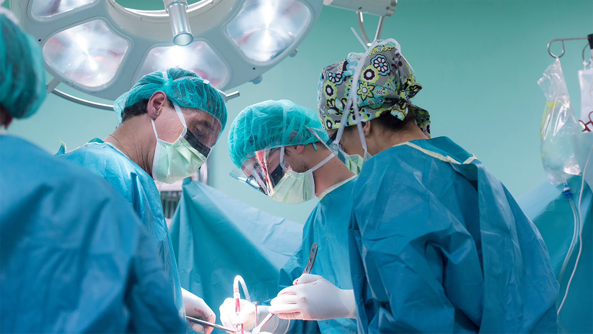
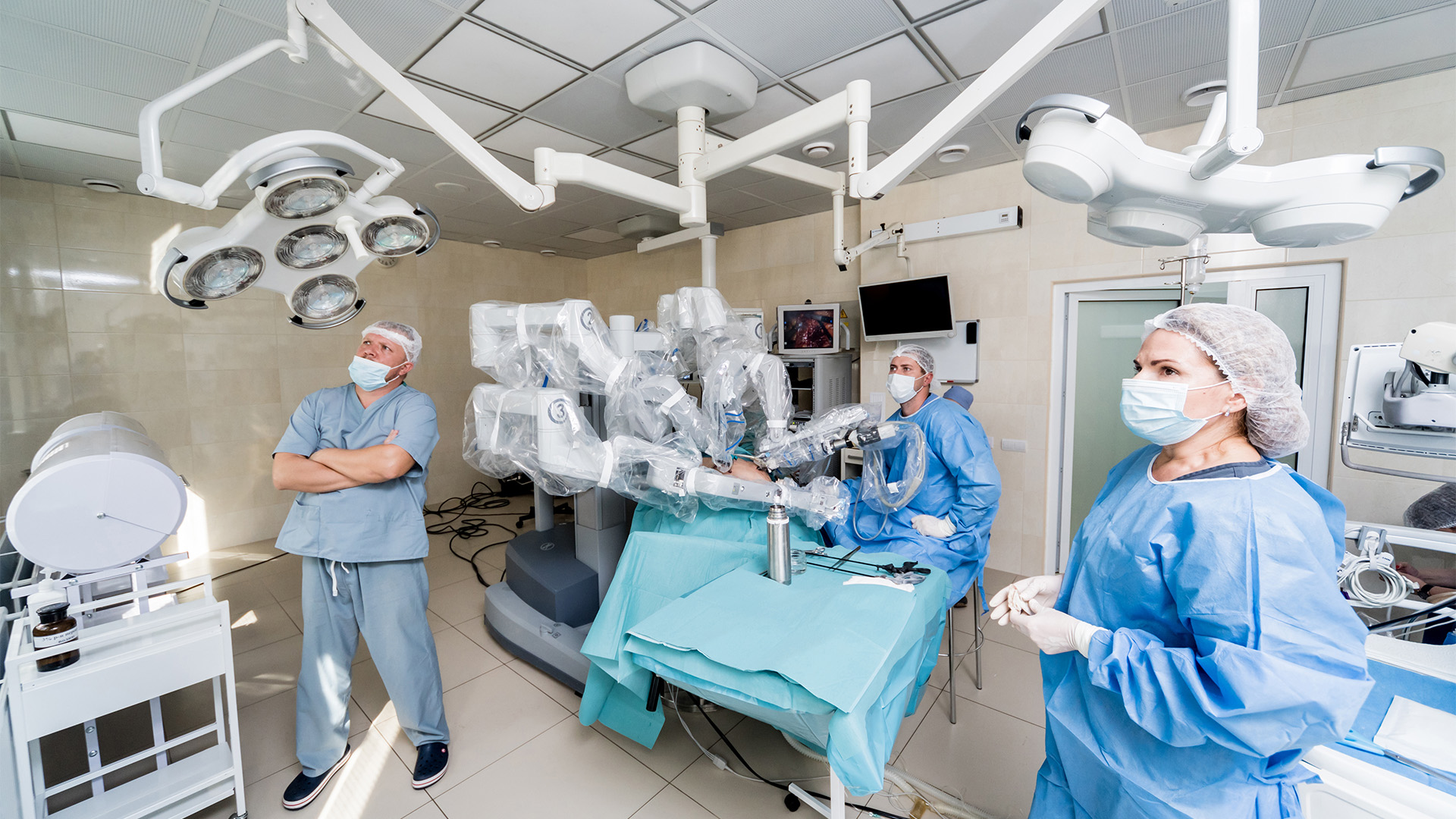
This video demonstrates the key surgical steps of transdiaphragmatic aorto-supra-celiac aortic extra-anatomic bypass. The case involved a 16-year-old boy who presented with shortness of breath on exertion. Echocardiography revealed type A interrupted aortic arch (IAA) with multiple collaterals draining into the distal part of the descending thoracic aorta (DTA), generalized wall hypokinesia, and an ejection fraction (EF) of 35 percent.
After the induction of general anesthesia, a midline sternotomy and upper midline laparotomy were performed. The peritoneum was opened, the left lobe of the liver was retracted and the stomach was mobilized to expose the diaphragmatic crura. The esophagus was dissected and looped.
The supraceliac aorta was approached by incising the median arcuate ligament, and the aorta was dissected and looped. The supraceliac part of the aorta was found to be small in caliber.
The pericardium was opened over the ascending aorta, while the pericardium over the right atrium (RA) was kept intact. The right pleura was opened, and a transdiaphragmatic rent was made anterior to the inferior vena cava (IVC) to create adequate space. A 12 mm collagen impregnated double-velour Dacron graft was tunneled through this diaphragmatic rent into the right pleural cavity.
Heparin was administered, and a Satinsky side-clamp was applied over the supraceliac part of the aorta. An end-to-side anastomosis of the Dacron graft with the supraceliac part of the aorta was performed using 4-0 Prolene sutures. After confirming adequate flow through the proximal end of the Dacron graft, the proximal anastomosis was fashioned onto the distal part of the ascending aorta under partial side-clamp control. Pressures were kept under control while the anastomosis was performed.
The position of the graft was confirmed below the diaphragm to ensure that there were no kinks. In this case, the graft was routed transdiaphragmatically and laid within the right pleural cavity making direct contact only against the liver surface. It was not in contact with any hollow viscera, such as the stomach or esophagus. Notably, secondary aortoenteric fistulas most commonly develop due to erosion into adjacent hollow organs. The liver, being a solid organ, carries a substantially lower risk of fistula formation.
Additionally, in the referenced series describing similar extra-anatomic reconstructions, no specific preventive measures against visceral adhesion were reported. The proximal anastomosis was performed using 4-0 polypropylene sutures. Adequate hemostasis was achieved.
References
- Messner G, Reul GJ, Flamm SD, Gregoric ID, Opfermann UT. Interrupted aortic arch in an adult single-stage extra-anatomic repair. Tex Heart Inst J. 2002;29(2):118-121.
Disclaimer
The information and views presented on CTSNet.org represent the views of the authors and contributors of the material and not of CTSNet. Please review our full disclaimer page here.
 402 views on Vimeo
 748 views