


Chest Wall

This video demonstrates a robotic-assisted resection of the left fifth rib for oligometastatic melanoma involving the chest wall. An 83-year-old man with a history of cutaneous melanoma presented with progressive left posterior chest wall pain...


This prospective European Society of Thoracic Surgeons (ESTS) Drainology Collaborative registry study examined chest-drain practice after pulmonary resection in 3,348 patients across 21 international centers, 88 percent of whom underwent...
This prospective cohort is the largest of its kind involving 240 pediatric patients enrolled over eight years and followed for a median of 80 weeks. Patients were screened with a...

A 63-year-old woman presented with myasthenia crisis and ptosis, for which she was treated with immunoglobulin. Her medical history included type 2 diabetes mellitus (DM), hypertension, and hypothyroidism. A computed...

This interview centers on a research paper titled “Mortality Risk and Cause of Death Associated With Removal of the Adult Thymus: Analysis of the U.S. Thymoma Population.” At the 2026...
In this month’s episode of The Atrium, Dr. Alice Copperwheat explores chest drains with Leanne Ashrafian, thoracic surgeon and CTSNet Senior Editor. They discuss patient considerations, equipment used, positioning of...
CTSNet Editor-in-Chief Joel Dunning spoke with Dr. Dawn Jaroszewski about optimizing pectus surgery. Dr. Jaroszewski addressed key issues, amounts of bars used in the procedure, and suppressing displacement of bars....
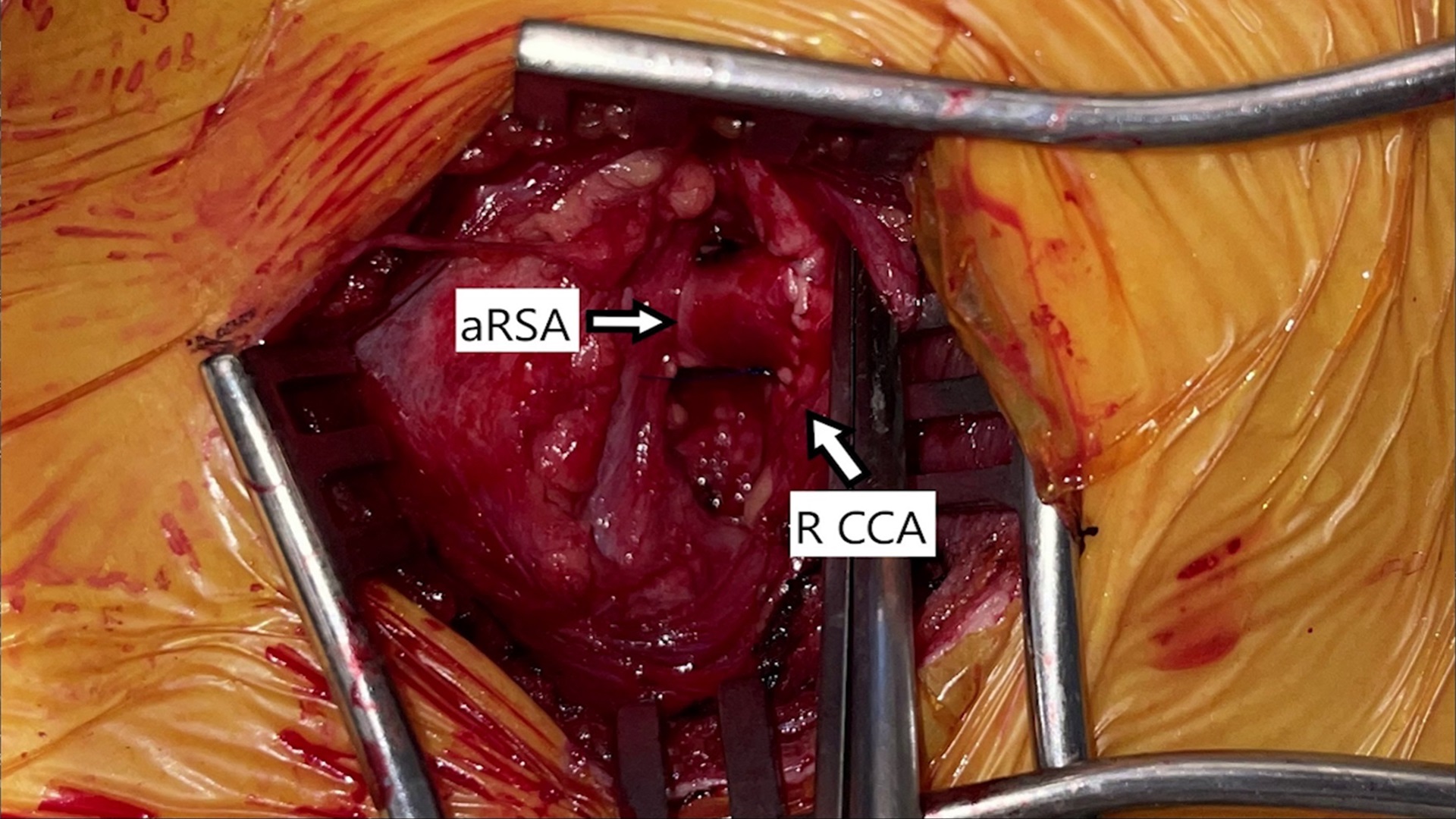
The patient was a 27-year-old female with a history of postural orthostatic tachycardia syndrome and Ehlers Danlos syndrome who presented with dysphagia to solids since 2021. Her work up was...
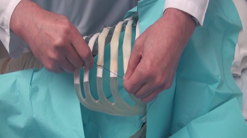
After chest wall resection, large defects are created in the bony structure of the chest. To preserve the integrity of the chest wall, a reconstruction procedure is often required. Multiple...



This is a Ravitch procedure that was modified to maximize the relief of cardiac compression. The usual version of the Ravitch operation involves leaving the lateral ribs in situ, and...


First, note that there are many accepted strategies for wound closure. The technique will vary based on the weight of the patient and complexity of the wound. Regardless of the...
 …
…This video presents the case of the third redo slide tracheoplasty and esophagoplasty following a button battery ingestion in a previously healthy fifteen-month-old child. The patient presented to the emergency...

At the 2024 STS Annual Meeting, CTSNet Editor in Chief Joel Dunning and CTSNet senior editors set out to interview the most knowledgeable CT surgeons from around the world. Stay...