Diaphragmatic defects involving the central tendon are uncommon and may present diagnostic uncertainty, particularly when identified incidentally on imaging. Surgical management can be technically challenging due to attenuated tissue quality and close proximity to vital structures, including the liver and inferior vena cava.
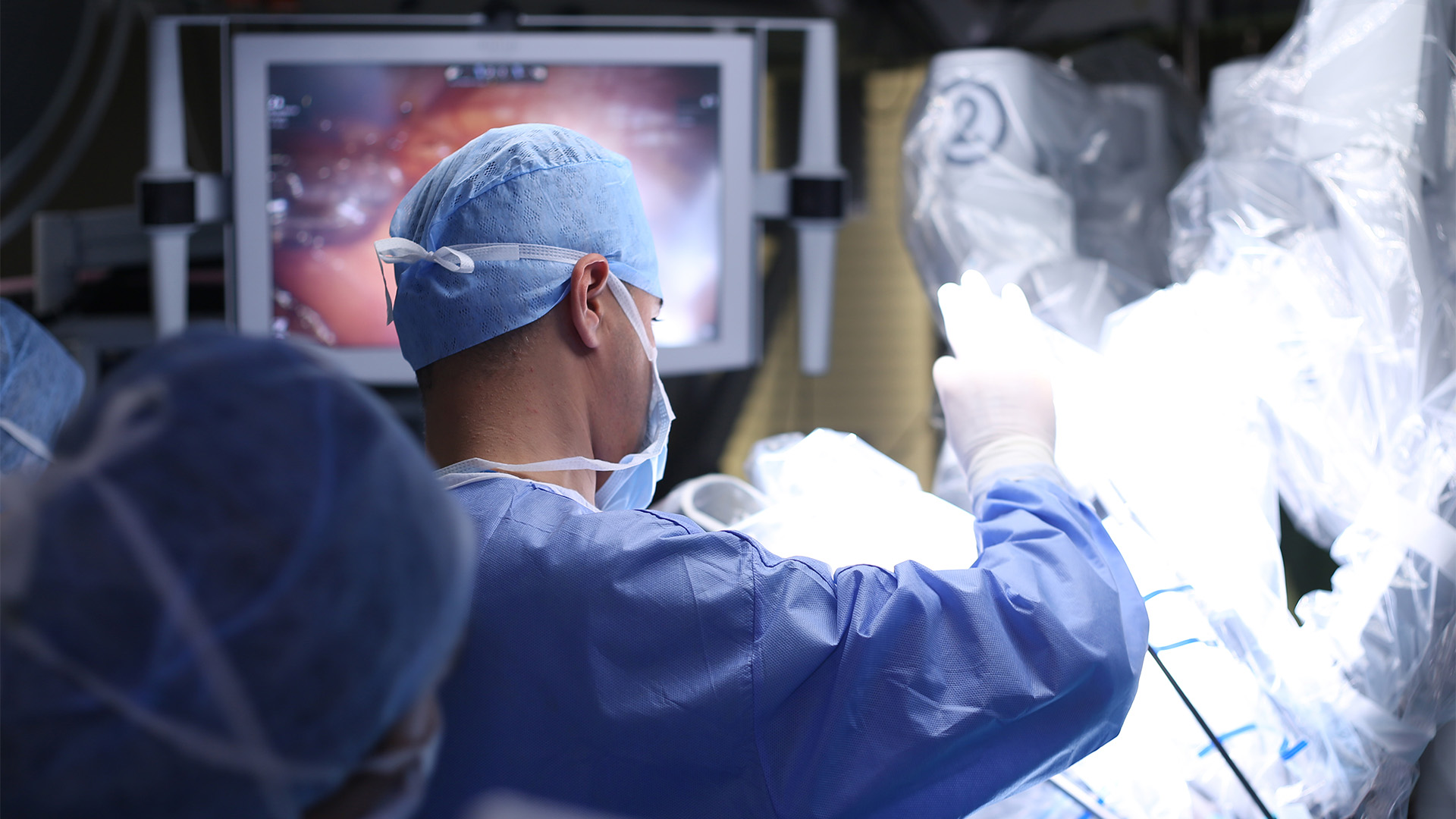
The authors present a video case report with intraoperative footage of a patient undergoing robotic repair of diaphragmatic herniation through a fenestrated central tendon identified on preoperative imaging. Intraoperatively, multiple fenestrations within the central tendon were identified, with herniation of the hepatic dome into the thoracic cavity. The video demonstrates the use of a robotic platform to facilitate safe dissection, assessment of tissue integrity, and tailored repair.
The diaphragmatic defect was managed using a combination of primary closure and reinforced suturing techniques to accommodate the attenuated central tendon while minimizing the risk of hepatic injury and caval distortion. The patient tolerated the procedure well, with an uncomplicated postoperative course and chest drain removal on postoperative day one.
Robotic repair of diaphragmatic herniation through a fenestrated central tendon represents a safe and effective approach in selected patients, offering enhanced visualization and suturing precision in anatomically constrained regions where conventional thoracoscopic techniques may be limited.
Disclaimer
The information and views presented on CTSNet.org represent the views of the authors and contributors of the material and not of CTSNet. Please review our full disclaimer page here.
 170 views on Vimeo