Skip to content
Skip to content

The 6th Annual Innovation Video Competition Is Now Accepting Entries!

READ MORE
Not a member yet? Register now.
  • ABOUT
    • Leadership
    • Participation
    • Featured Sponsors
    • Submission Guidelines
    • Competitions
      • Innovation Video Competition
    • Community
      • Volunteer
      • Badges
    • Contact Us
  • EXPLORE CTSNET
    • CARDIAC
    • THORACIC
    • CONGENITAL
    • Clinical Care Professionals
    • JANS
    • Events
    • Journals
  • Videos
    • Atlas of Cardiac Surgery
    • Webinars
    • Master Class
    • Guest Interviews
    • Conference Sessions
  • Podcasts
    • The Beat With Joel Dunning
    • The Atrium
    • The Cardiac Recovery Room
    • The Lifeline
  • Profiles
    • Users Directory
    • Organizations
  • CAREERS
    • US Career Center
    • European Career Center
  • ABOUT
    • Leadership
    • Participation
    • Featured Sponsors
    • Submission Guidelines
    • Competitions
      • Innovation Video Competition
    • Community
      • Volunteer
      • Badges
    • Contact Us
  • EXPLORE CTSNET
    • CARDIAC
    • THORACIC
    • CONGENITAL
    • Clinical Care Professionals
    • JANS
    • Events
    • Journals
  • Videos
    • Atlas of Cardiac Surgery
    • Webinars
    • Master Class
    • Guest Interviews
    • Conference Sessions
  • Podcasts
    • The Beat With Joel Dunning
    • The Atrium
    • The Cardiac Recovery Room
    • The Lifeline
  • Profiles
    • Users Directory
    • Organizations
  • CAREERS
    • US Career Center
    • European Career Center
SUBMIT CONTENT
DONATE
JOIN

welcomes you to the new, improved website!

READ MORE
  • ABOUT
    • Leadership
    • Participation
    • Featured Sponsors
    • Submission Guidelines
    • Competitions
      • Innovation Video Competition
    • Community
      • Volunteer
      • Badges
    • Contact Us
  • EXPLORE CTSNET
    • CARDIAC
    • THORACIC
    • CONGENITAL
    • Clinical Care Professionals
    • JANS
    • Events
    • Journals
  • Videos
    • Atlas of Cardiac Surgery
    • Webinars
    • Master Class
    • Guest Interviews
    • Conference Sessions
  • Podcasts
    • The Beat With Joel Dunning
    • The Atrium
    • The Cardiac Recovery Room
    • The Lifeline
  • Profiles
    • Users Directory
    • Organizations
  • CAREERS
    • US Career Center
    • European Career Center
Not a member yet? Register now.
  • ABOUT
    • Leadership
    • Participation
    • Featured Sponsors
    • Submission Guidelines
    • Competitions
      • Innovation Video Competition
    • Community
      • Volunteer
      • Badges
    • Contact Us
  • EXPLORE CTSNET
    • CARDIAC
    • THORACIC
    • CONGENITAL
    • Clinical Care Professionals
    • JANS
    • Events
    • Journals
  • Videos
    • Atlas of Cardiac Surgery
    • Webinars
    • Master Class
    • Guest Interviews
    • Conference Sessions
  • Podcasts
    • The Beat With Joel Dunning
    • The Atrium
    • The Cardiac Recovery Room
    • The Lifeline
  • Profiles
    • Users Directory
    • Organizations
  • CAREERS
    • US Career Center
    • European Career Center
  • ABOUT
    • Leadership
    • Participation
    • Featured Sponsors
    • Submission Guidelines
    • Competitions
      • Innovation Video Competition
    • Community
      • Volunteer
      • Badges
    • Contact Us
  • EXPLORE CTSNET
    • CARDIAC
    • THORACIC
    • CONGENITAL
    • Clinical Care Professionals
    • JANS
    • Events
    • Journals
  • Videos
    • Atlas of Cardiac Surgery
    • Webinars
    • Master Class
    • Guest Interviews
    • Conference Sessions
  • Podcasts
    • The Beat With Joel Dunning
    • The Atrium
    • The Cardiac Recovery Room
    • The Lifeline
  • Profiles
    • Users Directory
    • Organizations
  • CAREERS
    • US Career Center
    • European Career Center
SUBMIT CONTENT
DONATE
JOIN
You are here:
  1. Robotic & Endoscopic Cardiac

Robotic Diaphragm Plication—Technical Pearls

  • November 8, 2023

By: Benjamin Palleiko, Samantha Kaliszewski, Feiran Lou, Karl Fabian Uy, Mark Maxfield

Keywords:

  • Thoracic
  • Robotic & Endoscopic Cardiac

This video demonstrates a robotic diaphragm plication with a focus on technique and technical pearls.

The patient is a seventy-two-year-old woman with a history of phrenic nerve paralysis secondary to surgical division during a mediastinal mass resection in 1999. Following this operation, she had persistent dyspnea on exertion and worsened when supine. In 2020, she was diagnosed with COVID-19 requiring hospitalization, after which her dyspnea symptoms significantly worsened. Preoperative imaging demonstrated an elevated left hemidiaphragm. She underwent pulmonary function testing with an FEV1 of 52 percent and DLCO of 57 percent. A preoperative transthoracic echocardiogram showed normal biventricular systolic function and an ejection fraction of 55 percent. Because of her worsening dyspnea symptoms, elevated hemidiaphragm from phrenic nerve paralysis, and no clear cardiopulmonary cause of her dyspnea, she was recommended to undergo robotic diaphragm plication.

The Surgery

The operation was performed via a left-sided transthoracic approach. However, this patient had a history of chest radiation and sternotomy, and in the case of frozen chest, this operation could have been performed through an abdominal approach.

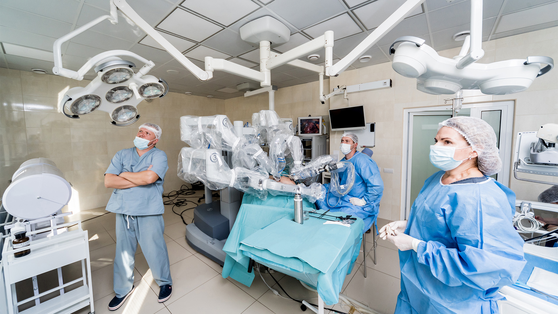
For the room setup, the robot normally comes in from the patient’s right for a standard lung resection. For diaphragm plication, however, it was set to thoracic left so that the boom could swing inferiorly instead of superiorly. A double lumen was placed, as was an NG tube that to suction and minimize dilation of the stomach. An 8 mm camera port was placed in the midline, superiorly. This is usually in the fourth intercostal space. Next, a 12 mm assistant port was placed posterior and inferior in approximately the eighth intercostal space. The anterior and posterior 8 mm ports were placed in line with the camera port. The posterior port should be in line with the camera port, posterior to the scapula, and if it is put too low, suturing will be difficult. 

A retraction port was then placed posterior and superior, in line with the camera port, and was used to retract the lung superiorly. A thirty-degree scope was used for port placement and a complete intercostal nerve block with Exparel, after which a zero-degree scope was used. The operation begins with a tip-up grasper in arm one, Cadiere forceps in arm two, a zero-degree scope in arm three, and a long bipolar grasper in arm four.

With insufflation set to 8 mmHg, the floppy and redundant diaphragm was visualized. The tip-up in the posterior port retracted the lobe superiorly with a cigar. Classically, a plication was performed from anteromedial to posterolateral to ensure that the diaphragm was lined up. Parallel lines were then made in the diaphragm to guide suture placement.

The first stitch was a barbed suture, which facilitates placing the first stitch under tension. All subsequent sutures were 0-Ethibond cut to 18 cm. A CT-2 needle was used for both the barbed and 0-Ethibond sutures.

Next, the diaphragm was grasped with the Cadiere forceps and pulled upward to avoid suturing the stomach or colon on the left and liver on the right. The initial suture was placed at the midpoint of the diaphragm using a Mega SutureCut needle driver in arm four.

The medial sutures tend to be more technically challenging. If the ports for two and four are placed too far inferior, then the sutures in this area can be technically difficult to place. Small bleeding from the diaphragm is to be expected and almost always resolves on its own. In this case, a slip knot was used to tie the sutures under tension. The sutures were placed to the full width of the diaphragm. The sutures on the posterolateral diaphragm were easier to place.

The insufflation was then turned off to assess diaphragm movement. After a satisfactory diaphragm plication, the diaphragm should be taut, as could be seen in this case. A 24 Fr Smart drain was then placed and the lung was inflated. 

Postoperatively, the patient’s pain was well controlled on oral medications and her chest tube was removed on postoperative day one. A postoperative day two chest X-ray showed the left hemidiaphragm in a more anatomically appropriate position, though still with slight elevation. The patient was discharged home on postoperative day two.

On postoperative day eighteen, the patient was seen in clinic where she was recovering well, with slow improvement in her breathing and activities. Notably, her dyspnea when supine had improved. However, she did have a chest X-ray done that showed a moderate sized pleural effusion. She underwent thoracentesis in clinic with 600 cc of simple appearing serosanguinous fluid removed. A subsequent chest X-ray demonstrated decreased size of the effusion and improved aeration of the lung. She was sent home on Lasix for one week with plans for a repeat chest X-ray in three weeks. Two months postoperatively, the patient’s breathing had significantly improved and her chest X-ray showed a small, stable, likely reactive pleural effusion. She will continue to follow up with thoracic surgery.


Disclaimer

The information and views presented on CTSNet.org represent the views of the authors and contributors of the material and not of CTSNet. Please review our full disclaimer page here.

Add Comments Cancel reply

You must be logged in to post a comment.

Not a member yet? Register now.
Vimeo 1041 views on Vimeo

CITATION

Palleiko BA, Kaliszewski S, Lou F, Uy K, W. Maxfield M. Robotic Diaphragm Plication—Technical Pearls. November 2023. doi:10.25373/ctsnet.24529792
DOI https://doi.org/10.25373/ctsnet.24529792
TAGS
  • Robotic Thoracic Surgery

Related Content

Article

Register for the Inaugural ‘In Africa for Africa’ Cardio-Thoracic Conference in Cape Town!

By: Marianna Mast

Podcast

The Beat With Joel Dunning Ep. 168: Humanoid Robots and AI in Surgery

By: Joel DunningGold ContributorSilver Commentor

Video

Robotic-Assisted Biventricular Durable Assist Device Implantation: The First In Human Experience

By: Feras Khaliel

JANS Article

Humanoid Robots in the Operating Room: A Framework for Staged Integration of Embodied AI in Surgery

Author(s): Aamir Amin

JANS Article

Total Endovascular Repair of Ascending Aortic Pathology: The ENDOASC Multicentre Global Study

Author(s): Vincent SierSilver Contributor

Video

Management of SVC Injury During Right VATS Thymectomy for Myasthenia Gravis

By: Jakraphan Yu

Video

Single-Stage Repair of Left Ventricular Diverticulum with Ventricular Septal Defect Closure: A Video Illustration

By: Vatsal Vora

Video

3D Endoscopic Aortic Valve Replacement Combined With Extended Septal Myectomy via Right Lateral Minithoracotomy

By: Glib Yemets

Video

Transaxillary Direct-View Double Valve Surgery: A Step-by-Step Procedural Guide

By: Michele Galeazzi

Video

Aorto-Mitral Angle Determines Ease of Mitral Valve Repair: Essential Anatomical Considerations

By: Jeevan Francis

233 S. Wacker Drive | Suite 4400
Chicago, Illinois 60606, USA

Linkedin X-twitter Facebook-f Youtube
  • Join CTSNet
  • Donate
  • Submit Content
  • Volunteer
  • Join CTSNet
  • Donate
  • Submit Content
  • Volunteer
  • Featured Partners
  • Careers
  • Press
  • Advertise on CTSNet
  • Featured Partners
  • Careers
  • Press
  • Advertise on CTSNet
  • CTSNet Policies
  • Privacy Policy
  • Disclaimer
  • Contact CTSNet
  • CTSNet Policies
  • Privacy Policy
  • Disclaimer
  • Contact CTSNet

233 S. Wacker Drive | Suite 4400
Chicago, Illinois 60606, USA

Instagram Linkedin X-twitter Facebook-f Youtube
  • Join CTSNet
  • Donate
  • Submit Content
  • Volunteer
  • Join CTSNet
  • Donate
  • Submit Content
  • Volunteer
  • Featured Partners
  • Careers
  • Press
  • Advertise on CTSNet
  • Featured Partners
  • Careers
  • Press
  • Advertise on CTSNet
  • CTSNet Policies
  • Privacy Policy
  • Disclaimer
  • Contact CTSNet
  • CTSNet Policies
  • Privacy Policy
  • Disclaimer
  • Contact CTSNet

233 S. Wacker Drive | Suite 4400
Chicago, Illinois 60606, USA

Instagram Linkedin X-twitter Facebook-f Youtube
  • Join CTSNet
  • Donate
  • Submit Content
  • Volunteer
  • CTSNet Policies
  • Privacy Policy
  • Join CTSNet
  • Donate
  • Submit Content
  • Volunteer
  • CTSNet Policies
  • Privacy Policy
  • Featured Partners
  • Careers
  • Press
  • Advertise on CTSNet
  • Disclaimer
  • Contact CTSNet
  • Featured Partners
  • Careers
  • Press
  • Advertise on CTSNet
  • Disclaimer
  • Contact CTSNet
  • Join CTSNet
  • Donate
  • Submit Content
  • Volunteer
  • Join CTSNet
  • Donate
  • Submit Content
  • Volunteer
  • Featured Partners
  • Careers
  • Press
  • Advertise on CTSNet
  • Featured Partners
  • Careers
  • Press
  • Advertise on CTSNet
  • CTSNet Policies
  • Privacy Policy
  • Disclaimer
  • Contact CTSNet
  • CTSNet Policies
  • Privacy Policy
  • Disclaimer
  • Contact CTSNet
© 2026 CTSNet Inc. All right reserved.
Go to Top