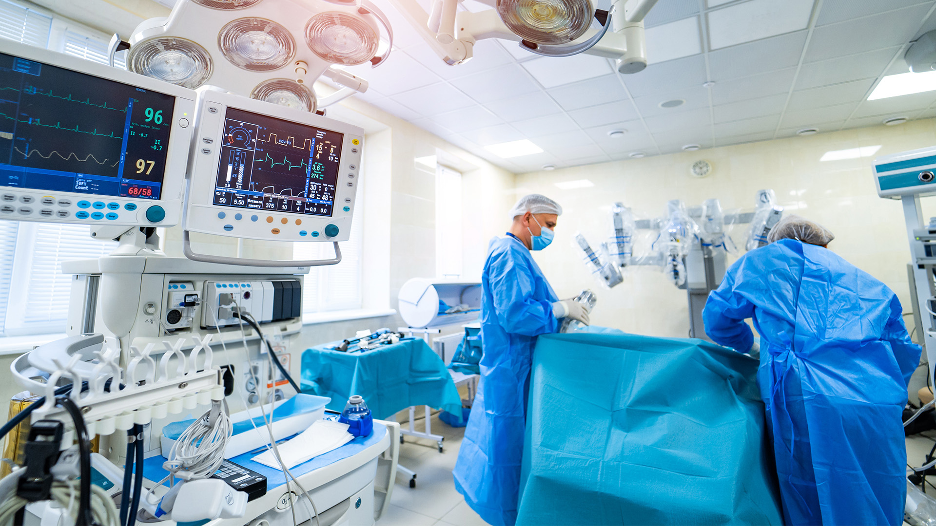
This clinical video demonstrates the preservation of a replaced left hepatic artery during the creation of a gastric conduit in a robotic-assisted minimally invasive esophagectomy (RAMIE) for post-neoadjuvant esophageal adenocarcinoma.
The patient was a 61-year-old male with cT3N0M0 (Stage IIA) lower-esophageal adenocarcinoma. He completed neoadjuvant therapy with four cycles of fluorouracil, leucovorin, oxaliplatin, and docetaxel. Post-induction imaging showed decreased fluorodeoxyglucose (FDG) uptake at the distal esophagus, which indicated a potential partial treatment response. He was taken to the operating room for a RAMIE.
The operation began with a standard five-port abdominal robotic setup, and immediate identification of aberrant arterial anatomy was made after the placement of the liver retractor. Careful dissection of the gastrohepatic ligament and celiac axis was performed to identify the vascular structures, which confirmed the presence of a replaced left hepatic artery (RLHA). Due to its large size, the decision was made to preserve it. All branches of the left gastric artery were identified and carefully ligated distal to the origin of the RLHA to maintain blood flow to the left lobe of the liver. Gastric mobilization and celiac lymphadenectomy were then carried out, followed by the creation of the gastric conduit. Pulsatility of the preserved RLHA was confirmed before the thoracic portion of the procedure was completed in the standard manner.
Aberrant left hepatic arterial (ALHA) anatomy, most commonly seen as an RLHA originating from the left gastric artery, is an important anatomical variant encountered in 5-10 percent of patients undergoing esophagectomy (1, 2). This case demonstrates the technical feasibility of ALHA preservation and offers a framework for managing this rare anatomical finding during esophagectomy. Furthermore, it highlights how the robotic approach’s improved visualization and instrument control facilitate arterial preservation without the need for converting to an open procedure (2).
This video is intended for both experienced thoracic surgeons and trainees aiming to enhance their knowledge and technical skills in managing this rare but potentially consequential anatomical variation. It emphasizes key perioperative decision-making, technical considerations for arterial preservation, and a framework for approaching aberrant vascular anatomy. The purpose of this video is to contribute to the limited esophagectomy-specific literature on ALHA and to underscore the feasibility and safety of arterial preservation, especially with the robotic approach.
This video submission is from the 2026 CTSNet Instructional Video Competition. Stay tuned to CTSNet.org and the CTSNet YouTube channel in the coming weeks to watch all entries from the competition, including the winning videos.
References
- Maki, H., Satodate, H., Satou, S., Nakajima, K., Nagao, A., Watanabe, K., Nara, S., Furushima, K., & Harihara, Y. (2018). Clinical evaluation of the aberrant left hepatic artery arising from the left gastric artery in esophagectomy. Surgical and Radiologic Anatomy, 40(7), 749–756. https://doi.org/10.1007/s00276-018-2022-4
- Hess, N. R., Rizk, N. P., Luketich, J. D., & Sarkaria, I. S. (2017). Preservation of replaced left hepatic artery during robotic-assisted minimally invasive esophagectomy: A case series. International Journal of Medical Robotics and Computer Assisted Surgery, 13(4). https://doi.org/10.1002/rcs.1802
Disclaimer
The information and views presented on CTSNet.org represent the views of the authors and contributors of the material and not of CTSNet. Please review our full disclaimer page here.
 72 views on Vimeo