


Anatomic segmentectomy has emerged as an effective option for the management of early-stage lung cancer; however, the division of intersegmental parenchyma remains technically challenging. The authors describe and illustrate the exclusion technique, a bronchus-last strategy that utilizes an endoleader catheter to guide parenchymal division and simplify segmentectomy.
Methods
The exclusion technique involves dissection and division of the segmental artery or arteries, followed by dissection and isolation of the segmental bronchus, which is intentionally left intact during parenchymal division.
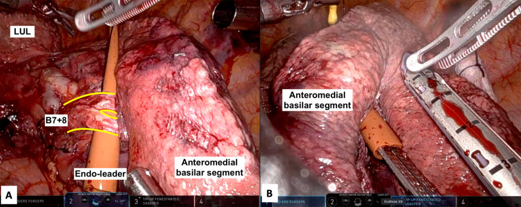
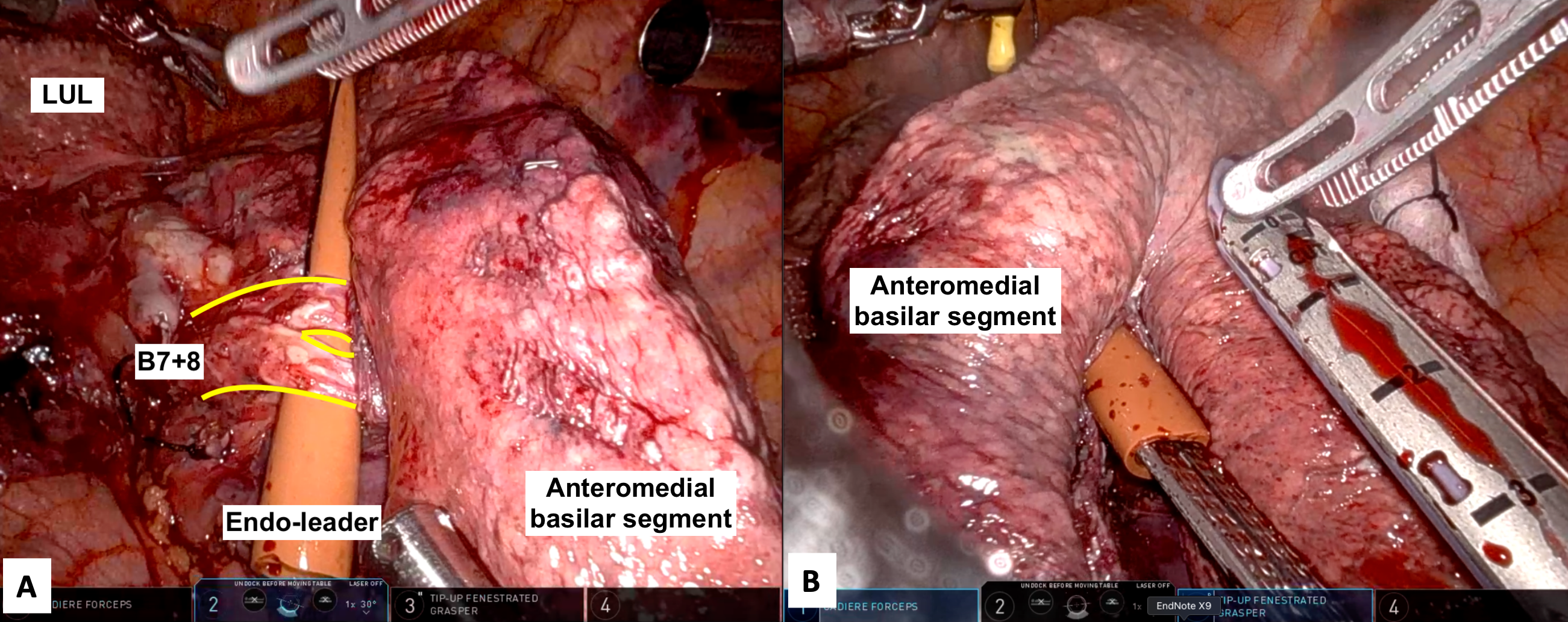
An endoleader catheter was positioned between the airway and the lung parenchyma, creating a tunnel to guide stapler passage (Figure 1). This technique is demonstrated across three robotic segmentectomy cases selected to highlight a range of anatomic challenges. In all cases, indocyanine green (ICG) was used following arterial division to demarcate the parenchyma prior to division. The segmental bronchus was divided last in each case to preserve orientation and facilitate safe, controlled dissection.
Results
Case 1
A right lower lobe composite basilar segmentectomy was performed. After division of the basilar pulmonary artery and vein, the endoleader was positioned between the basilar bronchus and lung parenchyma. ICG confirmed a clear boundary between the superior (S6) and the basilar segments (S7-10). Guided by the catheter, parenchymal stapling was performed without complication. Bronchial division completed the resection.
Case 2
A left lower lobe anteromedial basilar segmentectomy was performed in a patient with suspected interstitial lung disease. Following division of the segmental artery and vein and dissection of the bronchus, the exclusion technique was used to guide division of the parenchyma (Figure 1). Sequential stapling from the diaphragmatic surface upward was facilitated by the endoleader, which ensured proper stapler positioning despite poor visibility. Parenchymal division was completed safely, followed by bronchial division and confirmation of perfusion to the remaining lower lobe.
Case 3
A left upper lobe apical trisegmentectomy was conducted. The arterial branches supplying the apical trisegment were dissected and divided. The segmental airway was then dissected circumferentially and confirmed via bronchoscopy. After ICG demarcation, the endoleader was passed between the airway and parenchyma. Parenchymal division was performed using the exclusion technique, this time taking the segmental veins along with the parenchyma.
Conclusion
The exclusion technique offers an effective method for parenchymal division during robotic segmentectomy and is particularly useful in complex cases with deep segmental planes and challenging visualization. Key advantages include:
- Simplification of parenchymal division by using the intact segmental bronchus and the endoleader to create a guiding tunnel
- Protection of nearby vasculature from the stapler tip
- Optimized bronchial stump stapling
References
- Nomori H. Anatomical Segmentectomy for Lung Cancer. 1st ed. Springer; 2024. doi:https://doi.org/10.1007/978-981-99-5857-3
Disclaimer
The information and views presented on CTSNet.org represent the views of the authors and contributors of the material and not of CTSNet. Please review our full disclaimer page here.
 395 views on Vimeo












3 Comments
Interesting technique and great video! You perfectly explained the idea behind this approach. I could try this for my VATS segmentectomy as well. Thank you for sharing!
What is the yellow tip on your endolead catheter?
The yellow tip is a suture boot with a silk tie through the tip (secured by several knots on top of one another). The catheter is a red 10 Fr Rob-Nel PVC urethral catheter cut to 8-9 cm.