Anomalous aortic origin of a coronary artery from the opposite sinus is uncommon, and an interarterial course of an anomalous right coronary artery (RCA) carries a recognized risk of ischemia and sudden cardiac death.
When such a vessel also develops atherosclerotic disease, the abnormal ostial takeoff often makes percutaneous intervention difficult. Furthermore, proximal calcification may preclude unroofing or reimplantation, so bypass grafting was chosen in this case.
Isolated RCA disease is an unusual indication for minimally invasive surgery, and the right-sided approach is far less familiar than left-sided minimally invasive cardiac surgery (MICS) coronary artery bypass grafting (CABG).
Case Description
A 53-year-old woman with type 2 diabetes mellitus, hypertension, dyslipidemia, obesity (body mass index (BMI) of 31.2 kg/m2), and a fully recovered previous stroke presented with a non-ST-elevation acute coronary syndrome two months before surgery. Angiography showed no significant left main, left anterior descending or circumflex disease. The RCA originated from the left coronary cusp, with 70 percent diffuse calcified stenosis from the proximal to the mid vessel, and computed tomography angiography supported an interarterial course. Surgical revascularization was elected for single-vessel disease of an anomalous RCA from the opposite sinus.
Operative Technique
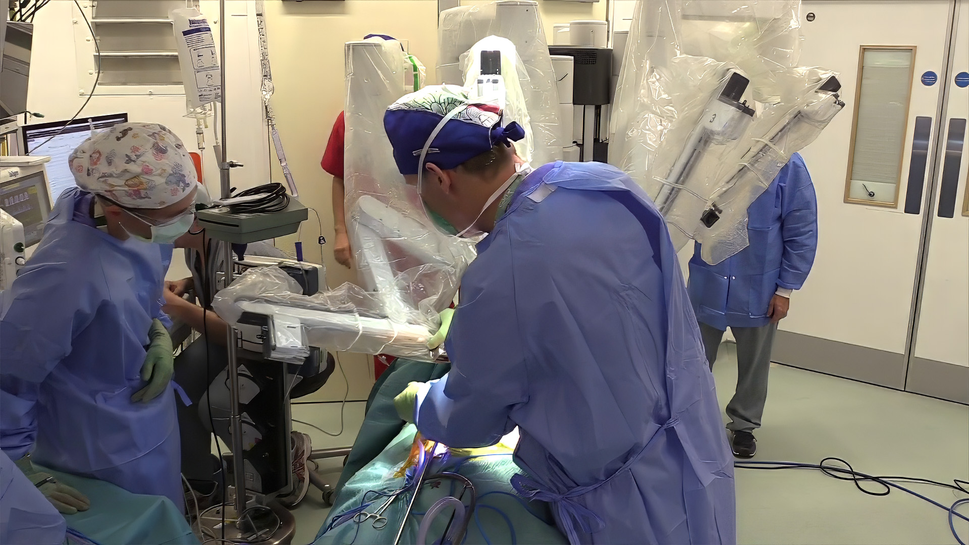
The patient was placed supine with the right chest mildly elevated. A double-lumen endotracheal tube was used to collapse the right lung. Three-port video-assisted thoracoscopy was used to harvest the right internal mammary artery (RIMA) in a skeletonized fashion through a 10 mm camera port and two 5 mm working ports positioned so that instruments approach the vessel along its axis. A right fourth intercostal space anterior minithoracotomy was made for the anastomosis. The pericardium was opened anterior to the phrenic nerve and suspended to bring the mid RCA into the wound. The mid RCA was then stabilized with an epicardial stabilizer and controlled with a vessel loop. An arteriotomy was made distal to the calcified segment, a 1.75 mm intracoronary shunt was inserted, and the RIMA was anastomosed end-to-side with running 7-0 polypropylene off pump.
Result
Operative time was 3 hours and 45 minutes, with 200 mL of blood loss, without conversion to sternotomy or cardiopulmonary bypass. The patient was extubated at eight hours and discharged on postoperative day five. At two weeks, she was free of chest pain and ambulating independently, with well-healed incisions.
Discussion
Right-sided MICS off-pump coronary artery bypass (OPCAB) is a reasonable option for isolated right coronary disease, particularly when the anatomy makes percutaneous intervention, unroofing, and reimplantation unappealing.
Three requirements differ from left-sided MICS CABG. First, port placement must be planned for a mirror-image field; ports set as for a left-sided harvest leave the surgeon working across the pedicle. Second, the mid RCA sits deeper in epicardial fat than the left anterior descending artery (LAD), and pericardial suspension is what brings the anastomosis within reach of a small incision. Third, single-lung ventilation is essential to maintain the working space during harvest.
Avoiding sternotomy and cardiopulmonary bypass is attractive in a patient with diabetes, obesity, and previous stroke, in whom sternal wound complications and neurologic injury are the main concerns. As a single case, this video documents feasibility and technique. Mid-term patency data is needed.
References
- CTSNetVideo. RA Right-Sided Minimally Invasive Coronary Artery Bypass for Anomalous Origin of the RCA. Published online April 6, 2026. Accessed September 11, 2026. https://www.youtube.com/watch?v=N8rKIo8h9yI
- Gräni C, Kaufmann PA, Windecker S, Buechel RR. Diagnosis and Management of Anomalous Coronary Arteries with a Malignant Course. Interv Cardiol. 2019 May 21;14(2):83-88. doi: 10.15420/icr.2019.1.1. PMID: 31178934; PMCID: PMC6545977.
Disclaimer
The information and views presented on CTSNet.org represent the views of the authors and contributors of the material and not of CTSNet. Please review our full disclaimer page here.
 2 views on Vimeo