
ALERT!
This site is not optimized for Internet Explorer 8 (or older).
Please upgrade to a newer version of Internet Explorer or use an alternate browser such as Chrome or Firefox.
An Approach to Modified Bedside Percutaneous Tracheostomy During the COVID-19 Pandemic
Wiesel O, Brichkov I, Shaw J. An Approach to Modified Bedside Percutaneous Tracheostomy During the COVID-19 Pandemic. June 2020. doi:10.25373/ctsnet.12459821
Background
The COVID-19 pandemic has resulted in a rapid increase in patients requiring mechanical ventilation. For patients who survive the acute phase of infection, tracheostomy may prevent long-term sequelae of prolonged endotracheal intubation, conserve sedative and paralytic medications, and facilitate endotracheal suctioning, weaning from mechanical ventilation, and cohorting of patients in a designated weaning unit. However, the risk of spreading the COVID-19 virus through aerosolizing procedures such as tracheostomy is well documented (1). Standards for safe practice when performing tracheostomy under these circumstances are not yet established.
The authors describe their protocol for the performance of tracheostomy in COVID-19-infected patients with special attention to procedural technique, personal protective equipment (PPE), and minimization of participating personnel. In their experience at a tertiary care, community-based teaching hospital, their protocol allowed them to perform 10-15 tracheostomies per week with a small dedicated team in a manner designed to minimize risk to both patients and healthcare workers under COVID-19 surge conditions.
Setting
The authors perform tracheostomies in an ICU setting, preferably in rooms with negative pressure capability. Where open spaces have been converted into COVID-19 units, they use portable HEPA filters. To standardize patient selection, they formed an institutional advisory team composed of intensive care specialists, pulmonologists, surgeons, and palliative care consultants for careful consideration of individualized timing, risks, and benefits.
Patient Selection
The ideal timing of tracheostomy for COVID-19-infected patients remains undefined. Due to severe hypoxia and lung injury, the authors found that many patients are not candidates for early tracheostomy (2). The authors recommend performing the procedure at approximately 2-3 weeks, once the patient has recovered from the initial cytokine storm, and ventilator parameters have stabilized and are adequate to perform tracheostomy.
Preparation
The designated team should train in appropriate donning and doffing of PPE. Each operator dons an N95 mask, head and foot coverings, a powered air-purifying respirator (PAPR), orthopedic hood, impermeable gown, and two pairs of gloves (Image 1). The help of an assistant and ample disinfectant wipes during donning and doffing are recommended according to the CDC guidelines (3). To minimize donning and doffing, the authors scheduled consecutive tracheostomies within a single unit to allow performance of successive procedures, with operators changing only the outer protective gown, hood, and gloves between procedures.
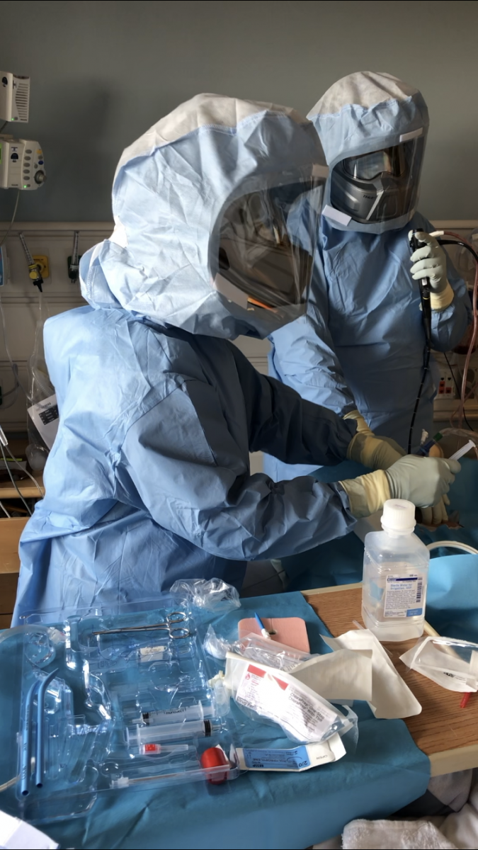
Image 1
Prior to initiation of the procedure, the intensivist, anesthesiologist, and surgeons assess again if the patient will be able to tolerate ventilator changes during the procedure. After initial set-up and positioning, the assisting nurse and/or resident is dismissed. The ideal team includes two attending surgeons (bronchoscopist and primary operator) and a senior anesthesiologist (equipped with a PAPR). A series of simple visual hand signals prompt the anesthesiologist to pause and resume ventilation as needed.
Procedural technique
The different techniques of tracheostomy are described elsewhere (4-6). To reduce risk of contamination and maximize the team’s safety, the authors modified our technique further as follows:
- They added PAPR to the standard CDC recommended PPE (3).
- The ventilator circuit is inspected for leaks or loose connections.
- If available, disposable bronchoscopes are used.
- The patient should be paralyzed to mitigate cough and aerosolization during procedure.
- Once the patient is adequately sedated, paralyzed, and pre-oxygenated, they recommend introducing the bronchoscope into the oropharynx between the vocal cords parallel to the endotracheal tube.
- With closed suctioning, the endotracheal tube is advanced distally under bronchoscopic guidance with the balloon inflated.
- Based on anatomical landmarks, the needle is introduced percutaneously into the trachea and the guide wire is then placed, followed by serial dilations.
- After the last dilation, ventilation is temporarily halted, and only then an 8.0 cuffed Shiley tracheostomy is passed into the lumen while the endotracheal tube is simultaneously withdrawn under bronchoscopic visualization to the level of the tracheostomy. Once the tracheostomy tube is confirmed in place with end tidal CO2, the endotracheal tube is removed.
- Ventilation is resumed once the tracheotomy tube cuff has been inflated and the tracheostomy is connected to the ventilator.
- Closed in-line suctioning is recommended to check for hemostasis; therapeutic bronchoscopy with aspiration of the distal airway should be avoided unless absolutely necessary.
- In the majority of the cases, the side-by-side technique was successfully completed with placement of an 8.0 endotracheal tube. Glottic or vocal cord edema may make passing the scope parallel to the endotracheal tube difficult, in which case a standard percutaneous technique might be employed under apnea or the procedure can be aborted and converted to open at a later time.
Discussion
The authors’ protocol for tracheostomy during the COVID-19 pandemic was designed to facilitate patient flow from ICU to dedicated weaning units, and maximize the safety of healthcare workers performing this high-risk airway procedure. While bedside tracheostomy offers the advantage of eliminating viral transmission in the operating room or during transport, the traditional bronchoscopic guided procedure increases the chance of virus aerosolization. While they speculate that the modified side-by-side bronchoscopic technique reduces aerosolization, it by no means eliminates it. Their approach, which maximizes healthcare worker PPE, limits donning and doffing, and minimizes personnel, which has allowed them to perform multiple procedures efficiently in multiple ICU settings, while avoiding high-risk patient transfer and operating room contamination. Institutional safeguards remain paramount to avert nosocomial transmission of COVID-19 during tracheostomy. Until then, the true risks of tracheostomy during the COVID-19 pandemic remains unknown.
References
- Wang W, Xu Y, Gao R, Lu R, Han K, Wu G, et al. Detection of SARS-CoV-2 in different types of clinical specimens. JAMA. 2020 Mar 11;323(18):1843-1844.
- https://www.entnet.org/content/tracheotomy-recommendations-during-covid-19-pandemic
- https://www.cdc.gov/coronavirus/2019-ncov/hcp/ppe-strategy/powered-air-purifying-respirators-strategy.html
- Ahmed N, Hare GM, Merkley J, Devlin R, Baker A. Open tracheostomy in a suspect severe acute respiratory syndrome (SARS) patient: brief technical communication. Can J Surg. 2005;48:6871.
- Byhahn C, Wilke HJ, Halbig S, Lischke V, Westphal K. Percutaneous tracheostomy: Ciaglia Blue Rhino versus the basic Ciaglia technique of percutaneous dilational tracheostomy. Anesth Analg. 2000;91:882–886.
- Angel L, Kon Z, Chang S, Rafeq S, Shekar SP, Mitzman B, et al. Novel percutaneous tracheostomy for critically ill patients with COVID-19. Ann Thorac Surg. 2020 April 25 [Epub ahead of print].
Disclosure
Jason Shaw and Igor Brichkov received instructor's fees from Cook Medical.
Disclaimer
The information and views presented on CTSNet.org represent the views of the authors and contributors of the material and not of CTSNet. Please review our full disclaimer page here.


Comments