
ALERT!
This site is not optimized for Internet Explorer 8 (or older).
Please upgrade to a newer version of Internet Explorer or use an alternate browser such as Chrome or Firefox.
An Incidentally Found Sternal Foramen in a 16-Year-Old With Xiphodynia
History
A 16-year-old male with no past medical history initially presented for evaluation of xiphodynia. He reported that he had a prominent protrusion from the inferior aspect of his sternum, which caused him pain when lying prone and when diving during volleyball. A preoperative computed tomography (CT) scan incidentally revealed a 2.0 cm x 2.5 cm sternal foramen in the lower third of his sternum (figure 1). Given that there was evidence of healthy and intact surrounding cortical bone, sternal reconstruction was deferred, and he proceeded with xiphoid resection.
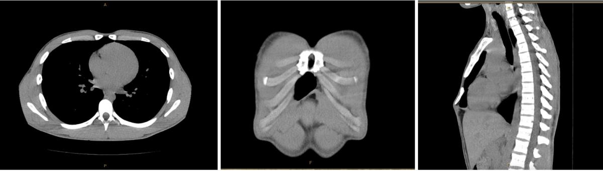
Figure 1: Computed tomography views of an inferior foramen.
Sternal Foramen
A sternal foramen (SF) is a common anatomical defect present in 2.5 to 13.8 percent of individuals (1). It arises from the incomplete fusion of the cartilaginous neonatal sternum. At six weeks of gestation, two longitudinal mesenchymal bands on the anterior chest wall begin to fuse at the midline. These bands, known as sternal bars, undergo fusion in a cranial to caudal direction until the 10th week of development (2). During the fifth and sixth months of gestation, the cartilaginous sternum begins to ossify in transverse segments, referred to as sternebrae. Contiguous sternebrae begin to fuse in the third and fourth years of life, and this process is usually completed by early adulthood (2). Any anomaly in this process can prevent proper fusion and may result in a sternal foramen, cleft, or other sternal defects.
Sternal foramina are typically asymptomatic; however, depending on their size, they can leave the vital underlying structures vulnerable to injury. This vulnerability may lead to catastrophic thoracic injury during acupuncture, chest surgery, or sternal biopsies. Approximately 11 to 20 percent of SFs lie directly against the pericardium, and more than 99 percent of all documented cases of sternal foramen are adjacent to the right ventricle (1). The aorta and lungs are also at risk of iatrogenic injury from a blind procedure. Therefore, thorough CT scans or sternal ultrasounds must be performed to rule out an SF before an initial procedure. However, such imaging is rarely performed outside of a hospital setting. Thus, acupuncture practitioners must have detailed knowledge of sternal anatomy, congenital anomalies, and proper oblique insertion techniques. In lean individuals with a skin-to-heart distance of 10-20 mm, there is still a risk of fatal cardiac tamponade despite the use of proper technique (2).
Management of Sternal Foramina
While surgical repair is indicated for sternal clefts and other severe thoracic malformations, there is no consensus on whether SFs require intervention. Thus, providers must consider factors such as the patient’s profession and body habitus, which may place them at increased risk of life-threatening injury when deciding between conservative or surgical management. Currently, there are no case reports detailing surgical repair of an SF. Instead, the consensus in the literature is that sternal foramina must be recognized early to avoid damaging underlying structures during other sternal interventions (4). In the case of severe sternal clefts, repair is indicated to protect the mediastinum and improve respiratory dynamics (5).
Operation
The patient was placed in a supine position on the operating table, and general anesthesia was induced. The prominent xiphoid process was palpated, and a 3 cm vertical midline incision was made. The soft tissue overlying the xiphoid process was divided with electrocautery. The muscular attachments on either side of the xiphoid, as well as the posterior diaphragmatic attachments, were freed bluntly and with electrocautery to the base of the xiphoid, taking care not to violate the pleura or peritoneum. Using a rongeur, the xiphoid process was resected in its entirety to the xiphisternal joint. The diaphragmatic muscle was reattached to the base of the sternum, covering the exposed left pleura. The posterior and anterior rectus sheaths were reapproximated, and the soft tissue was closed in layers to provide adequate coverage.
References
- Choi PJ, Iwanaga J, Tubbs RS. A Comprehensive Review of the Sternal Foramina and its Clinical Significance. Cureus. 2017;9(12):e1929. Published 2017 Dec 8. doi:10.7759/cureus.1929
- Babinski MA, de Lemos L, Babinski MSD, et al. Frequency of sternal foramen evaluated by MDCT: a minor variation of great relevance. Surg Radiol Anat. 2015;37(3):287-291. doi:10.1007/s00276-014-1339-x
- Garssen FP, Aalders MB, van der Poel MJ, Zuidema WP. Operative results after xiphoidectomy in patients with xiphodynia. Asian Cardiovasc Thorac Ann. 2021;29(8):779-783. doi:10.1177/02184923211019179
- Sungur EC, Tütün U, Tekin A. Incidentally Detected Sternal Foramen during an Urgent Surgical Revascularization: A Case Report. J Tehran Heart Cent. 2020;15(2):81-83. doi:10.18502/jthc.v15i2.4189
- Doğan R, Uysal S, Kumbasar U, Kesici S, Bayrakçı B. Surgical repair of a sternal cleft malformation. Turk Gogus Kalp Damar Cerrahisi Derg. 2019;27(4):597-600. Published 2019 Oct 23. doi:10.5606/tgkdc.dergisi.2019.18175
Disclaimer
The information and views presented on CTSNet.org represent the views of the authors and contributors of the material and not of CTSNet. Please review our full disclaimer page here.


