
ALERT!
This site is not optimized for Internet Explorer 8 (or older).
Please upgrade to a newer version of Internet Explorer or use an alternate browser such as Chrome or Firefox.
Situs Inversus Uniportal Video-Assisted Thoracoscopic Left Middle Lobectomy
By Diego Gonzalez Rivas, MD, Ricardo Fernandez, MD, Mercedes De La Torre, MD, and Lara Fontan, MD
Totalis Situs inversus (SIT) is an extremely rare condition. There are few reports in literature describing thoracic surgery in patients with this unusual anomaly. This video is the first report of a thoracoscopic lung resection of a pulmonary mass in a patient with SIT.
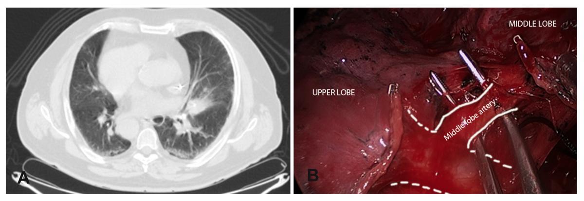
Clinical case: A 70 year-old male smoker was admitted to our department for surgery. He was diagnosed with SIT condition. A CT scan revealed a 3cm mass in the left middle lobe. A PET scan demonstrated uptake with no lymph node affection. The patient was proposed for video-assisted thoracoscopic (VATS) surgery.
Surgical technique:The patient was placed in a right lateral decubitus position. An uniportal video-assisted thoracoscopic approach through a single 4 cm incision was made in the left 5th intercostal space with no rib spreading. The left lung composed of three lobes and vascular and bronchial anatomy was exactly as a normal right side lung. Azygos vein, superior vena cava, and paratracheal space were mirror images of normal condition (Fig. 1A,B).

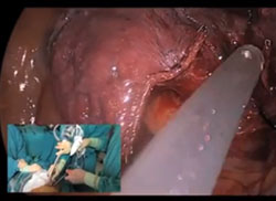
A 3cm mass was located in the middle lobe (Fig. 2A), too large and deep to perform a wedge resection so a middle lobectomy (Fig. 2B) with complete lymphadenectomy was performed (paratracheal and subcarinal space). A single chest tube was placed in the posterior part of the incision.
The total surgical time was 150 minutes. The patient was discharged on the second post-operative day. The final pathological examination revealed an organizing pneumonia.
Video:


Comments