
This video describes a surgical strategy for repairing interrupted aortic arch (IAA) associated with a common arterial trunk (CAT) in a 10-day-old baby (3.71 Kg). Echocardiography confirmed the diagnosis of IAA between the left carotid and left subclavian arteries and CAT type I. Truncal valve was quadri-leaflet with trivial central regurgitation.
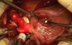
On the operating table, once the baby was put on cardiopulmonary bypass (CPB), cooled to 28 °C, and antegrade cerebral perfusion started, the duct was divided and its tissues entirely removed. The arch was repaired by using a reverse subclavian flap to reconstruct the superior part of the arch and a pulmonary homograft patch to repair the under surface of the arch. The cross clamp was then repositioned below the aortic arch and full CPB and rewarming were reinstituted. The ascending aorta was then separated from the pulmonary arteries without disconnecting them but using an "aortopulmonary window" type of repair with a Gore-Tex patch. The CAT repair was then continued in the usual fashion by exposing the VSD through a right ventriculotomy and closing it using a Dacron patch. A 12 mm Contegra conduit was then interposed between the right ventricle and the bifurcation of the pulmonary arteries. The patient was weaned from CPB in sinus rhythm and good haemodynamic stability.
In intensive care the baby was gradually weaned from inotropes and ventilation and discharged on the 4th post-operative day to the ward. The baby made good progress and was discharged from hospital 12 days post-operatively. At follow up, the baby remains asymptomatic, is gaining weight and without echocardiographic evidences of obstructions.


