
ALERT!
This site is not optimized for Internet Explorer 8 (or older).
Please upgrade to a newer version of Internet Explorer or use an alternate browser such as Chrome or Firefox.
Minimally Invasive Mitral Valve Replacement with a Robotic Digital Microscope
Cavozza C, Pellegrino P, Campanella A, et al. Minimally Invasive Mitral Valve Replacement with a Robotic Digital Microscope. January 2024. doi:10.25373/ctsnet.25112267
This video is part of CTSNet’s 2023 Innovation Video Competition. Watch all entries into the competition, including the winning videos.
Extracorporeal telescopes have been the latest addition to the neurosurgeons' armamentarium. The implementation of three-dimensional exoscopes in minimally invasive cardiac surgery seems feasible for safe utilization and may also be a promising tool in terms of surgical education. Understanding learning curves, informing surgical training, and evaluating procedures in practice are crucial for assessing a new surgical technique or technology.
A perfect image is essential in minimally invasive mitral valve surgery. The 3D exoscope allows for an enhanced image quality of the surgical field while also being more ergonomically favorable. The major benefits of the exoscope include the ocular independent visualization of the surgical field and the similar 3D visualization for all participants of the surgery (Fig 1).

Fig 1: The Aesculap Aeos® Robotic Digital Microscope combines the ergonomic and educational benefits typically found in “exoscopes” with the high-quality imaging.
The exoscope allows high-quality visualization of the operative field and an unobstructed view of the surgeon’s hands, instruments, and working angles (Fig 2). This allows a clear dual perception of the surgical field by the assisting surgeon and allows for a more efficient workflow thanks to robotic-assisted features along with remarkable vision quality. Assisted or automated positioning enables surgeons to execute precision movements through a multi-axis robotic arm, providing a 3D digital visualization display to enhance the precision needs that surgeons require for complex surgical procedures. It also includes a heads-up display that can be viewed simultaneously by all healthcare workers in the operating room with 3D glasses (Fig 3).
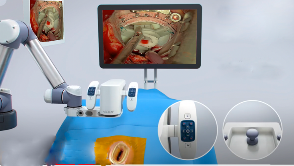
Fig 2: Assisted or automated positioning enables surgeons to execute precision movements, through a multi-axis robotic arm, providing a 3D 4K digital visualization display.
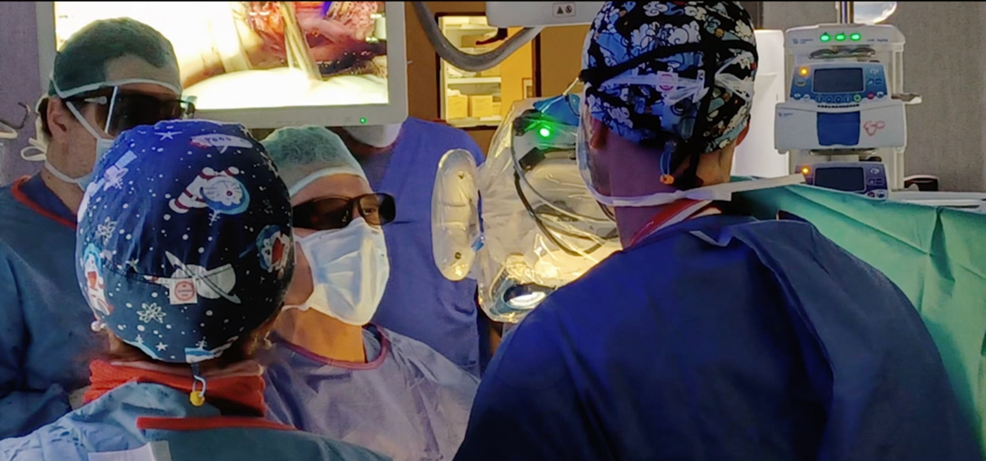
Fig 3: The heads-up display can be viewed simultaneously by all healthcare workers in the operating room with 3D glasses.
In addition, assisted or automated positioning enables surgeons to execute precision movements through a multi-axis robotic arm, providing a 3D 4K digital visualization display to enhance the precision needs that surgeons require for complex surgical procedures.
This video illustrates the exoscope technique. An eighty-year-old patient with severe chronic ischemic mitral regurgitation and impaired left ventricular function (ejection fraction < 40 percent) underwent mitral valve replacement. Functional MR was due to morphological and functional abnormalities of the left ventricle in which the main mechanism of MR was the asymmetric restriction of one posterior mitral leaflet.
Minimally invasive mitral valve surgery was performed as usual. The robotic platform allows easy and less invasive placement in the operating room (Fig 4).
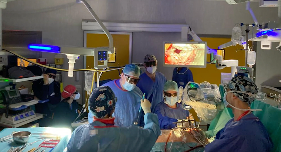
Fig 4: The robotic platform allows a easy and noninvasive placement into operating room.
Robotic assisted, precise repositioning places the camera head exactly, allowing for a more efficient workflow with a remarkable vision quality (Fig 5). Once optimal visualization is obtained, the mitral valve is inspected and surgery can be performed (Fig 6).
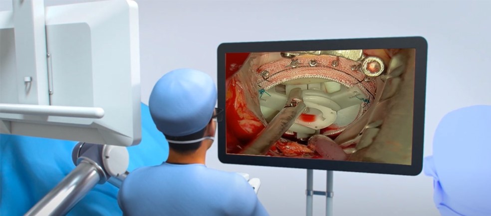
Fig 5: Robotic-assisted, precise repositioning places the camera head exactly, allowing for a more efficient workflow with a remarkable vision quality.
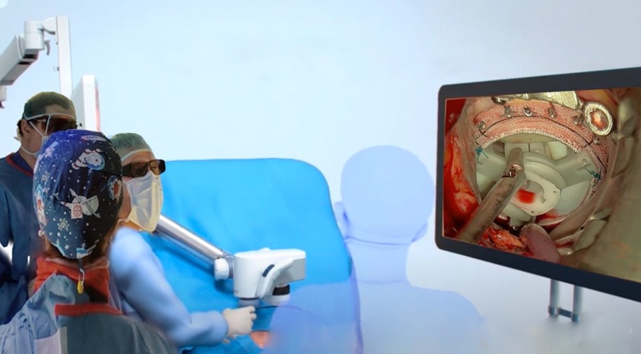
Fig 6: After optimal visualization is obtained, the mitral valve is then inspected and surgery can be performed as usual.
References
- Maurer, S.; Prinz, V.; Qasem, L.-E.; Lucia, K.E.; Rösler, J.; Picht, T.; Konczalla, J.; Czabanka, M. Evaluation of a Novel Three-Dimensional Robotic Digital Microscope (Aeos) in Neurosurgery. Cancers 2021, 13, 4273.
- Herlan S, Marquardt JS, Hirt B, Tatagiba M, Ebner FH. 3D Exoscope System in Neurosurgery-Comparison of a Standard Operating Microscope With a New 3D Exoscope in the Cadaver Lab. Oper Neurosurg (Hagerstown). 2019 Nov 1;17(5):518-524
- Langer DJ, White TG, Schulder M, Boockvar JA, Labib M, Lawton MT. Advances in intraoperative optics: a brief review of current exoscope platforms. Operative Surg. (2020) 19:84–93
- Haeren, Roel MD, PhD*,‡; Hafez, Ahmad MD, PhD*; Lehecka, Martin MD, PhD*. Visualization and Maneuverability Features of a Robotic Arm Three-Dimensional Exoscope and Operating Microscope for Clipping an Unruptured Intracranial Aneurysm: Video Comparison and Technical Evaluation. Operative Neurosurgery: January 2022 - Volume 22 - Issue 1 - p 28-34
- Hafez A, Haeren RHL, Dillmann J, Laakso A, Niemelä M, Lehecka M. Comparison of operating microscope and exoscope in a highly challenging experimental setting. World Neurosurg. 2021;147:e468-e475.
Disclaimer
The information and views presented on CTSNet.org represent the views of the authors and contributors of the material and not of CTSNet. Please review our full disclaimer page here.


