
ALERT!
This site is not optimized for Internet Explorer 8 (or older).
Please upgrade to a newer version of Internet Explorer or use an alternate browser such as Chrome or Firefox.
Pediatric Left VATS Upper Lobectomy for Infected Congenital Cystic Malformation
This video describes a left Video-Assisted Thoracoscopic (VATS) upper lobectomy in an 18-month old child with infected congenital cystic adenamatoid malformation (CCAM).
CCAM was diagnosed prenatally and then confirmed at birth by CXR and CT scan of the thorax which showed a large hyperinflated left upper lobe cyst with mediastinal shift to the right and compression of the lower lobe.
Parents refused surgery at birth and elected for regular follow up. At 18-months the child presented with sepsis secondary to infected cyst. This was initially treated with percutaneus drainage of the cyst and 6 weeks of antibiotics with complete resolution of the sepsis.
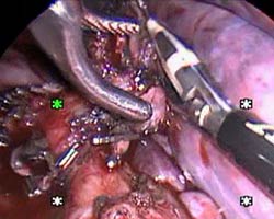
VATS lobectomy was performed electively. The patient was anaesthetized and single lung isolation was obtained by selectively blocking the left main stem bronchus. 3x5mm ports and one 15mm utility incisions were used. Dense chest wall adhesions from previous infection were taken down. Pulmonary arterial and venous branches to upper 1obe were doubly clipped and divided with an energy source device. The bronchus was stapled and divided with an endocutter. The lobe was removed through the utility incision.
Post-operative recovery was uneventful with full expansion of the lower lobe.


