
ALERT!
This site is not optimized for Internet Explorer 8 (or older).
Please upgrade to a newer version of Internet Explorer or use an alternate browser such as Chrome or Firefox.
Right VATS Pneumonectomy
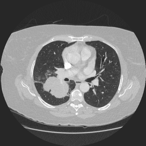
Figure 1: Preoperative CT scan
In this case, the preoperative CT demonstrated a 6 cm central tumor crossing the oblique fissure between the upper and lower lobes. The case was discussed at the authors’ tumor board, and a right pneumonectomy was recommended. Mediastinal nodes, sampled by EBUS/EUS, were negative.
The patient had severe adhesions between the lower lobe and the diaphragm. To avoid lung congestion, the veins and the truncus anterior were isolated first and then eventually divided. To avoid injury of the main pulmonary artery during dissection, particular care was taken to run the dissector on top of the right main bronchus. Eventually, the specimen was extracted in an endobag. All the steps of the procedure were completed without rib spreading, although the utility incision was slightly larger to allow extraction of the specimen.
The patient had minimal postoperative pain that was managed with a combination of Codeine and Paracetamol. The patient was discharged on the fourth postoperative day.


Comments