Content from the Journal of the American Neurological Surgeons.
The authors present the surgical management of four different late sequelae post-repair of tetralogy of Fallot with pulmonary atresia and criss-cross pulmonary arteries. The patient was an 11-year-old, 33.5 kg girl who was born with cyanosis due to pulmonary atresia and ventricular septal defect. She underwent an initial central shunt in the neonatal period, followed…
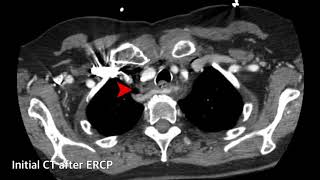
Perforation of Zenker’s diverticulum is a known risk of endoscopic procedures, and surgical transcervical repair or drainage is the standard management (1). Unresolved leak may require alternate means of drainage. An 82-year-old woman suffered esophageal perforation during attempted endoscopic retrograde cholangiopancreatography (ERCP). Following left transcervical exploration and drainage, the patient remained septic with free perforation…
Operation performed: Repair of type 1 common arterial trunk – bovine pericardial baffle (VSD patch) from inlet VSD to truncal valve and placement of 12 mm Hancock RV to PA Conduit. Repair of complete atrioventricular septal defect – Rastelli type C with 2 patch (VSD patch and ASD patch 1/primum patch) technique using bovine pericardium…
Mediastinitis represents an uncommon but potentially life-threatening complication following median sternotomy, with incidences ranging from 0.25-5% (1). It may result from direct hematogenous spread of bacteria or from the extension of infection from adjacent structures e.g., esophagus, airways, lungs, or head and neck (descending necrotizing infection). Diagnostic criteria have been described by the Centers for…
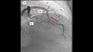
The authors present an interesting cases of a myocardial bridging as a cause of exertional chest pain. This is the case is of a 42-year-old woman with a past medical history of HTN and PAHTN who has been worked up by cardiology for chest pain with minimal exertion. The CXR didn’t show any acute abnormalities,…
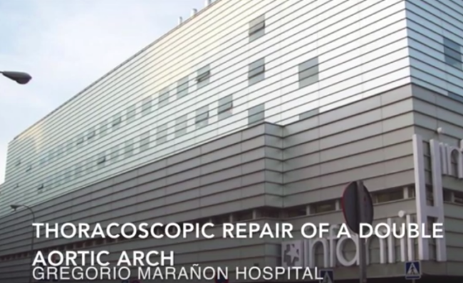
Introduction Double aortic arch is a relatively rare cardiovascular malformation. It is an anomaly of the aortic arch in which two aortic arches form a complete vascular ring that can compress the trachea and/or esophagus. Case Report The patient was a 13-year-old, 77 kilogram girl with solid dysphagia. The CT scan revealed a double aortic…
These foregut-derived congenital malformations are usually discovered prenatally on ultrasound and can be seen at any point along the tracheobronchial tree. So you might be thinking to yourself, what’s the dilemma? Well, when it comes to adults, bronchogenic cysts can be seen throughout the chest and abdomen, but, in general, they rarely occur and even…
Dr. Marc Gerdisch is the chief of cardiovascular and thoracic surgery and surgical director of the Heart Valve Center at Franciscan Health, Indianapolis, Indiana. He is also a senior partner with the Illinois-based Cardiac Surgery Associates specializing in complex heart valve surgery, as well as an associate clinical professor of thoracic and cardiovascular surgery at…
The authors present a video case report of an ex-premature, corrected age of 35 weeks who was referred because of a large patent ductus arteriosus, heart failure, and failure to thrive. After multiple unsuccessful attempts at patent ductus arteriosus device closure, it spontaneously closed. At some point during the procedure, the catheter wire was noted…
The patient was a 21-year-old man with a history of worsening cough, chest pain, and increasing chest wall asymmetry during the previous six months. Imaging demonstrated a large heterogeneous mass of the left hemithorax, causing compression of the heart, pulmonary artery, left lung, and displacing the mediastinum to the right. Alpha-fetoprotein and beta-HCG were increased.…
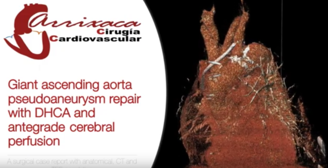
Aortic ascending pseudoaneurysm (AAP) is a rare and challenging pathology whose understanding could be demanding for cardiothoracic residents. Video reports of this condition are scarce and the correlation between the surgical findings and the preoperative diagnostic images are often not performed. The authors present a clinical case which focuses on the correlation between anatomy and…
The authors present a case of successful minimally invasive esophagectomy after prior open Nissen fundoplication and gastrostomy, not previously described in the literature. A 43-year-old man presented with dysphagia to solids only. He had a past medical history of reactive airway disease and known Barrett’s esophagus, for which he was getting endoscopy surveillance every three…
The authors present a case of a transgastric laparoscopic-assisted endoscopic resection in a 64-year-old man with an incidentally found 5.4 cm gastric GIST. The mass was located endoscopically near the gastroesophageal junction. Three transgastric ports were placed and the stomach was insufflated. The mass was excised with ultrasonic shears and removed endoscopically to avoid enlarging…
A medical student interested in surgery broadly, and cardiothoracic surgery specifically, interviews a fellow who completed a general surgery residency and is in his last year of a cardiothoracic fellowship. The interview covers the residency and fellowship process, his daily life, and why others should consider cardiothoracic surgery. Disclaimer The information and views presented on…
This is a case of the use of the Artisential VATS Wristed Instruments and also an automated camera holder bringing together two excellent technologies to simulate as closely as possible a robotic lobectomy. The authors performed a middle lobectomy on a woman with stents who continued clopidogrel and aspirin throughout the perioperative course. The advantages…
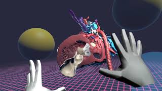
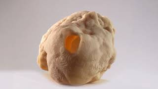
A patient with extremely low ejection fraction, pseudoaneurysm of the left ventricle free wall with a volume exceeding volume of the left ventricle, and secondary mitral regurgitation due to postinfarction papillary muscle dislocation was admitted to the hospital. Before the operation, based on CT scans, a 3D model of the left ventricle with an aneurysm…


 welcomes you to the new, improved website!