Content from the Journal of the American Neurological Surgeons.
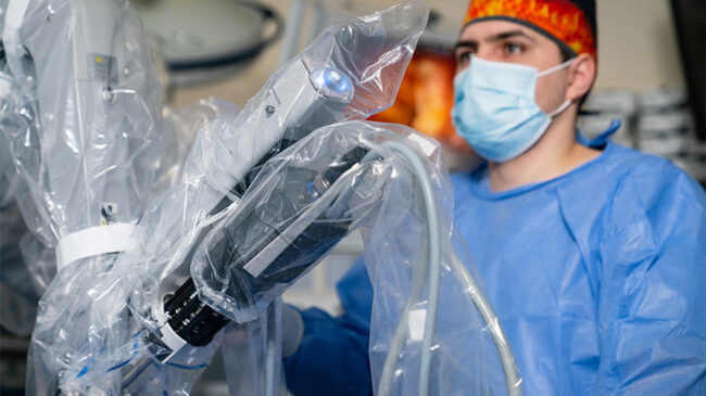
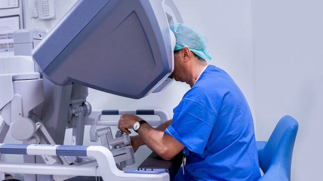
The authors report a redo robotic-assisted mitral valve replacement (MVR) in a 50-year-old male with prior bioprosthetic MVR via sternotomy, presenting with New York Heart Association (NYHA) class III dyspnea and significant prosthetic mitral inflow obstruction (mean/peak gradient 10/20 mmHg) with preserved biventricular function. The procedure was performed using a robotic platform with del Nido cardioplegia for myocardial protection, and a mechanical prosthesis…
This clinical video provides a detailed, step-by-step overview of the robotic-assisted convergent (ROK AF) procedure for the treatment of atrial fibrillation. The technique combines minimally invasive epicardial ablation with endocardial catheter-based ablation to achieve durable rhythm control, particularly in patients with persistent or long-standing persistent atrial fibrillation. This how-to video is designed to serve as a practical guide…
In this video, the authors present the case of a 65-year-old female diagnosed with a central squamous cell carcinoma of the left upper lobe with nodal involvement in stations 5 and 6, initially detected during the evaluation of a persistent cough. Computed tomography revealed a 57 mm centrally located mass in the left upper lobe with involvement…
This video demonstrates a robotic left lower lobectomy using bedside stapling to improve efficiency and assist with the rapid flow of the operative procedure. Interestingly, with the Si system, this was the routine way of performing the operation, but with the advent of robotic stapling, interest in bedside stapling has declined. However, the advantages of bedside stapling are numerous. First, with more junior surgeons or trainees, if the senior surgeon is scrubbed, they can significantly speed the cadence of the operation by…
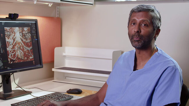
The 79-year-old male patient had a transcatheter aortic valve replacement (TAVR) placed at another institution in another state, although he presented with more complex issues than only aortic stenosis. The patient was a retired internist who originally had a bicuspid valve with aortic regurgitation (AR) and aortic stenosis (AS). A transcatheter 29 mm valve was placed. However, he was subsequently admitted with acute heart failure due to acute mitral valve regurgitation and anemia. Additionally, he had aortic insufficiency, and the team was concerned about hemolysis. He underwent a TAVR computed tomography (CT) scan in addition to a transesophageal echocardiogram (TEE) to clarify his diagnosis. The 3Mension program was used…
A gentleman in his 60s with advanced oesophageal cancer (cT4bN1M0) and extensive nodal disease developed a malignant tracheoesophageal fistula (TEF). Due to the presence of the fistula, he was not a candidate for radiotherapy given the high risk of exacerbating the defect. He completed five cycles of 5-fluorouracil (5-FU)-based chemotherapy and was on maintenance pembrolizumab administered every six weeks. Clinically, he…
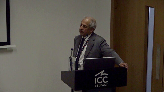
At the Society for Cardiothoracic Surgery in Great Britain and Ireland Annual Meeting, leading surgeons presented on various cardiac topics, including mitral annular calcification (MAC), mitral regurgitation (MR), reverse remodeling in ischemic mitral regurgitation (IMR), and several rapid-fire case debates. Dr. Gianluca Lucchese, a consultant cardiac surgeon at Guy’s and St Thomas’ NHS Foundation Trust in London, UK, and a senior clinical lecturer at King’s College London, delivered a presentation titled “The Big MAC: Contemporary Treatment Strategies and the…
This video presents a case of an older patient with severe pectus who had only one Nuss bar. Over four months, the bar fractured the thin sternum and migrated over it, exiting the thoracic cavity. This video demonstrates this complication, the removal of the bar, and how the surgeons subsequently performed the Ravitch procedure, and using two Stryker Variax plates to elevate the sternum and stabilize the fracture. Disclaimer The information and views presented on CTSNet.org represent the views of the authors and contributors of the material…
Background Congenital absence of the midline sternum is a rare chest wall anomaly that exposes mediastinal structures and causes paradoxical respiration, leading to respiratory and feeding difficulties. This report describes the successful surgical management of an infant with complete absence of the midline sternum using a chondral flap technique to restore stable native tissue coverage. Case Description…
Introduction Video-assisted thoracoscopic thymectomy (VATS) is an established approach for myasthenia gravis. The authors present a bilateral VATS thymectomy, highlighting key technical aspects. Case Description A 29-year-old male with ptosis was diagnosed with myasthenia gravis. Positron emission tomography (PET) imaging showed a 2 × 2 cm fluorodeoxyglucose (FDG)-avid anterior mediastinal lesion (standardized uptake value [SUV] 4.69). After medical optimization, bilateral VATS thymectomy was performed, starting…
Traumatic tracheobronchial injury is a rare but life-threatening complication of blunt chest trauma, accounting for approximately 0.5-2 percent of cases (1). These injuries most commonly occur near the right main bronchus due to the fixed position of the carina and reduced mediastinal protection (2). Prompt diagnosis and surgical intervention are essential to prevent respiratory failure and long-term complications. This video demonstrates the case…
This video demonstrates a patient in their 50s who experienced shortness of breath. Twenty years ago, they underwent pectus and breast implants to hide the defect, but now they require a Nuss procedure to anatomically correct the defect. First, the pectus implant was removed, and the incision was used to perform chondrectomies to help lift the sternum adequately, which is usually more challenging in a 50-year-old patient due to chest stiffness. The breast implants were also removed, one of which had already ruptured and was causing pain. Following this, a Prime Pectus Nuss procedure was performed with three bars,…
This video showcases a Nuss procedure performed after a previous Ravitch operation. The patient had undergone the Ravitch procedure 10 years prior, but the condition had recurred, leading to significant shortness of breath. The technique for the Nuss repair is demonstrated using the following seven golden rules: Elevate the chest Open the mediastinum widely Use…
At the Society for Cardiothoracic Surgery in Great Britain and Ireland Annual Meeting, leading surgeons presented on various thoracic topics, including pectus care, chest wall resection and reconstruction, and sympathectomy. Dr. Joel Dunning, CTSNet Editor-in-Chief and a cardiothoracic surgeon at James Cook University Hospital, Middlesbrough, UK, delivered a presentation on “Pectus Care in 2026.” He…
In this top-notch interview, CTSNet Editor-in-Chief Joel Dunning spoke with Dr. Andrea Steely, an Assistant Professor of Cardiac Surgery in the Division of Cardiothoracic Surgery at the University of Utah Health, Salt Lake City, UT, USA, about aortic surgery and long-term patient follow-up. They discussed the critical importance of educating both patients and surgeons about aortic disease, and the most effective strategies for follow-up care after aortic surgery. The conversation also covered testing genetic…
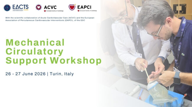
Join Us in Turin in June for This Interactive MCS Workshop! Providing didactic teaching, hands on dry lab operative experience, and practical demonstrations, this MCS Workshop welcomes everyone involved in heart failure care. Surgeons, anesthesiologists, perfusionists, acute care specialists, interventional cardiologists and heart failure nurses—all who participate in the management of critically ill patients…