Content from the Journal of the American Neurological Surgeons.
It is always preferable to save as much lung parenchyma as possible during tracheobronchial surgeries, if possible, keeping in mind the importance of complete disease clearance (R0 resection) to avoid recurrences. Previously, pneumonectomies were performed in most cases with tumors in the main bronchus. Now whenever possible, lung sparing surgeries are the norm. With more…
This video demonstrates a robotic left upper lobectomy in a woman with a history of smoking, COPD, and breast cancer treated with surgery and adjuvant radiotherapy-hormone therapy. The particularity of this patient is that she presented with complete situs inversus (three lobes and two pulmonary fissures as in a right-sided lung). She had a suspicious…
At the 2024 AATS Annual Meeting, CTSNet senior editors set out to interview the most knowledgeable CT surgeons from around the world. Stay tuned for more interviews from the meeting. CTSNet Senior Editor Mara B. Antonoff spoke with Hyun Koo Kim about single-port robotic-assisted thoracic surgery. Dr. Kim addressed the advantages of single-port robotic-assisted surgery compared to…
The authors present a case of a fenestrated atrial septal defect (ASD) closure in a 6-year-old female child with severe pectus excavatum and pulmonary hypertension, performed via vertical right axillary thoracotomy. A 6-centimeter vertical skin incision was made in the right mid axillary line extending from the second to the fourth intercostal spaces, and the…
In this CTSNet original webinar, Senior Editor and thoracic surgeon Dr. Leanne Ashrafian led a panel discussion with physicians who have devopled a novel approach to advanced endometriosis treatment: “A Multidisciplinary Approach to Treating Complex Endometriosis.” The webinar was broadcasted to a live audience on July 25, 2024. The multidisciplinary panel included Dr. Shaheen Khazali, Consultant Gynecological…
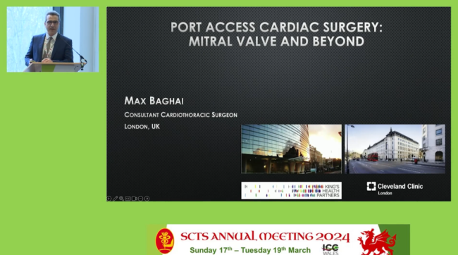
In March 2024, the Society for Cardiothoracic Surgery in Great Britain and Ireland brought together surgeons from around the world to discuss CT surgery topics and share knowledge. Watch for more SCTS 2024 videos in the coming weeks. At the Society for Cardiothoracic Surgery Annual Meeting 2024, Max Baghai delivered a speech on port-access…
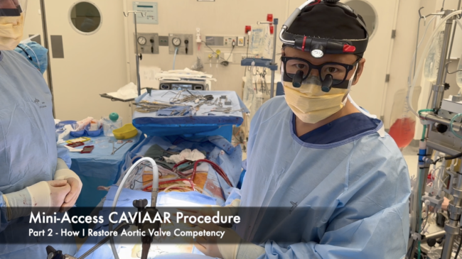
Aortic root remodelling with external subaortic annuloplasty (CAVIAAR procedure) enables physiological movements of the aortic cusps within three symmetrically reconstructed neosinuses, thus preserving root expansibility with the interleaflet triangles (1). In addition, the external aortic annuloplasty prevents annular base dilatation. In Part 2, the author demonstrated how to restore the aortic valve competency, by creating…
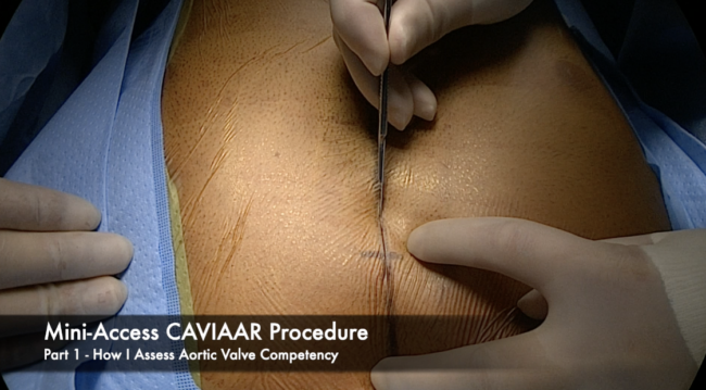
Aortic root remodeling with external subaortic annuloplasty (CAVIAAR procedure) enables physiological movements of the aortic cusps within three symmetrically reconstructed neosinuses, thus preserving root expansibility with the interleaflet triangles (1). In addition, the external aortic annuloplasty prevents annular base dilatation. In Part 1, the author demonstrated how to assess the aortic valve competency by using…
This is a Ravitch procedure that was modified to maximize the relief of cardiac compression. The usual version of the Ravitch operation involves leaving the lateral ribs in situ, and then passing a bar or mesh between them to lift the sternum. However, this often leaves the ribs pressing against the heart and does not…
This video details a thoracic surgical procedure in which a video-assisted thoracoscopic surgery (VATS) right basilar segmentectomy is performed on a patient with a typical carcinoid tumor. The procedure is enhanced by using indocyanine green (ICG), a fluorescent dye inserted intravenously, that improves the visualization of anatomical structures. The video begins with a brief overview…
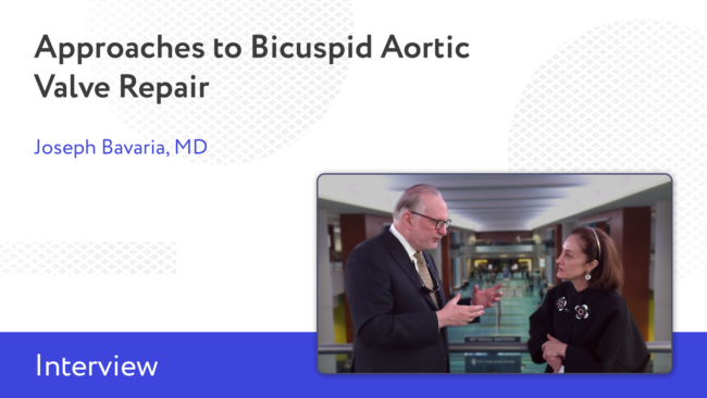
At the 2024 AATS Annual Meeting, CTSNet senior editors set out to interview the most knowledgeable CT surgeons from around the world. Stay tuned for more interviews from the meeting. CTSNet Board Member Marjan Jahangiri spoke with Dr. Joseph E. Bavaria about the different approaches to bicuspid aortic valve repair. Dr. Bavaria addressed tips for…
The authors present a case of a neoaortic valve repair with a HAART ring in a twenty-seven-year-old patient born with transposition of great arteries who underwent an arterial switch operation as a neonate. Indication for surgery was moderate-to-severe aortic insufficiency (AI), severe left ventricular (LV) dilation, and mild LV dysfunction. A redo median sternotomy was…
Surgically repairing pulmonary atresia is a challenging problem when there is complete absence of pulmonary arteries. To restore the continuity and confluence of the lung hila, a tubal structure is required. Although different types of biologic or nonbiologic materials have been used for this purpose, calcification, thrombosis, peeling formation by intimal hyperplasia, shrinkage, fibrosis,strictures, and…
In March 2024, the Society for Cardiothoracic Surgery in Great Britain and Ireland brought together surgeons from around the world to discuss CT surgery topics and share knowledge. Watch for more SCTS 2024 videos in the coming weeks. At the 2024 SCTS Annual Conference, Aung Oo delivered a speech on AMDS and FET in acute…
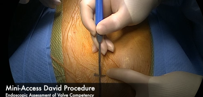
Valve sparing root replacement requires meticulous surgical techniques to restore aortic valve function. Several methods have been used to assess aortic valve competency post reconstruction, including visual inspection, water injection, and measurement of effective heights. However, it remains difficult to evaluate leaflet coaptation and degree of cusp prolapse when the aortic root is not pressurized.…
Introduction As the main source of cardiogenic emboli, left atrial appendage (LAA), accounts for more than 90 percent of embolic strokes (1, 2). It has been referred to as the most “lethal human attachment” (2). Various shapes, namely cactus, chicken wing, windsock, and cauliflower, have been described in the literature (3). These descriptions, however, have…