Content from the Journal of the American Neurological Surgeons.
This is a case of the use of the Artisential VATS Wristed Instruments and also an automated camera holder bringing together two excellent technologies to simulate as closely as possible a robotic lobectomy. The authors performed a middle lobectomy on a woman with stents who continued clopidogrel and aspirin throughout the perioperative course. The advantages…
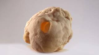
A patient with extremely low ejection fraction, pseudoaneurysm of the left ventricle free wall with a volume exceeding volume of the left ventricle, and secondary mitral regurgitation due to postinfarction papillary muscle dislocation was admitted to the hospital. Before the operation, based on CT scans, a 3D model of the left ventricle with an aneurysm…
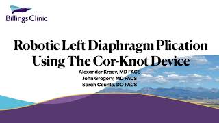
This video is a description of a novel technique using an automatic suture device (Cor-Knot) in robotic-assisted diaphragm plication. The slides go through the indications, OR set-up, operative steps, pitfalls, and pertinent points. The video is a demonstration of this technique. Disclaimer The information and views presented on CTSNet.org represent the views of the authors…
A 2-week-old, 3.5 kg child was transferred from a distant hospital (300 miles away) on congestive heart failure. Suspected diagnosis was ventricular septal defect (VSD), atrial septal defect (ASD), and pulmonary hypertension (PHT). On arrival at the authors’ NICU, the patient was stabilized with diuretics and vasodilators and eventually intubated. Echo showed an apical VSD…
This video demonstrates a case report of a 33-year-old man diagnosed with Tetralogy of Fallot (ToF). He had progressive exertional dyspnea with oxygen saturation 91% and NYHA class II-III. The preoperative echocardiogram showed large ventricular septal defect (VSD) with aortic overriding and right ventricular outflow tract (RVOT) obstruction. The operation plan was to perform total…
Atrial fibrillation (AF) is a major cause of structural heart disease, stroke, and increased hospitalizations. It has been estimated that 6-12 million people in the US will suffer from this condition by 2050 and 17.9 million people in Europe by 2060 (1). Management options by electrophysiologists include pharmacological therapy, cardioversions, and catheter ablations (2). Patients…
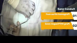
The authors present a stage I Yasui procedure in the form of a modified Norwood/Sano for a 2 kg, 6-day-old neonate with interrupted aortic arch type A, large posterior malalignment ventricular septal defect, and severely hypoplastic left ventricular outflow tract (Z-score = – 4.5). The goal was to achieve two-ventricle repair considering he had two…
Welcome to the sixth of twelve videos that compose the series “Thoracic Surgical Oncology – Technical Approaches,” by Professor Tristan D. Yan. To learn more about the series and see the complete list of videos, click here. This series of video tutorials demonstrates how to perform VATS lobectomy in a safe and simple way, using the…
The authors present a case of uniportal VATS left lower lobe radical lobectomy. The patient was a 44-year-old woman with breast cancer multimodal treated. The patient’s surgical history was represented by Madden mastectomy on the left side in 2019 and a left lower lobe wedge resection through minithoracotomy in 2020. The pathological report was lung…
The author reports the case of a vats left S8 segmentectomy. In this case, they used a coil to precisely localize the small lesion. The surgery began with pleural inspection, and then they started, as for any pulmonary oncological resection, by a radical mediastinal lymph node dissection. They moved next to the fissure, where they…
With this video, the authors present the case of a 53-year-old patient with synchronous NSCLC (adenocarcinoma) in the segments 2 and 9 of the right lung. The patient had limited lung function. The authors performed a uniportal VATS segmentectomy of segment 2 and 9 using Indocyanine green to define segmental margins. Histological analysis confirmed synchronous…
This video demonstrates a totally endoscopic transaortic extended septal myectomy for obstructive hypertrophic cardiomyopathy in an 81-year-old woman who was heavily symptomatic with class 3 shortness of breath. The LVOT peak gradient was measured 132 mm Hg on TOE. The authors used a totally endoscopic approach through a 3 cm, 2nd intercostal space (ICS), right…
The patient was a 6-year-old boy with no symptoms and a finding of a systolic heart murmur on the left sternal border on a routine examination. Echocardiogram showed left ventricular outflow tract obstruction by subaortic fibrous membrane with a peak gradient of 76 mm Hg and mild aortic valve regurgitation with preserved left ventricular function.…
Post-MI VSR repair by infarct exclusion technique was originally described by Cooley et al. has undergone many modifications and has been found to be a simple and reproducible technique with improved outcomes. The important advantages of this technique are: a) it maintains the geometry of the ventricle, b) it does not require reaction of the…
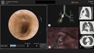
A 69-year-old man with chronic obstructive pulmonary disease and a 20-pack-year smoking history was found to have a 20 mm x 8 mm lung nodule concerning for malignancy in the periphery of the right lower lobe. A robotic-assisted bronchoscopy was performed to obtain a tissue diagnosis. The robotic bronchoscope consists of an inner bronchoscope (4.2…
Patient Selection Constrictive pericarditis results from a stiff pericardium that prevents adequate diastolic filling and can cause diastolic heart failure. The normal thickness of the pericardial is found in up to 18% of constrictive pericarditis cases (1). Myocardial and pericardial cells are both involved in the pathogenesis of pericarditis and constriction as indicated by the…